
Hydrocephalus simply means water in the brain. This is formed due to abnormality in the CSF circulation and formation. Due to this development of fluid occurs within the ventricles of brain and surrounding the brain.
1. Clinical features :
- Irritability, lethargy, poor appetite and vomiting.
- Enlargement of the head.
- The anterior fontanel is widely open and bulging.
- The scalp veins are dilated.
- Sunset sign – The forehead seems to be broad and eyes might deviated downward.
- Brisk-tendon reflex, spasticity, clonus and Babinsky’s sign.
2. Causes :
| Congenital Malformations | Acquired Causes |
| 1. Aqueduct obstruction 2. Dandy-Walker syndrome 3. Congenital central nervous system infections 4. Craniofacial anomalies 5. Benign intracranial cysts 6. Arnold Chiari malformation 7. Vein of Galen aneurysms | 1. Inflammation – Meningitis, granulomatous conditions/sarcoid. 2. Absorption blockage – Intracranial hemorrhage 3. Tumors and cysts – In the Posterior fossa, pineal, 3rd ventricle. Like colloid cyst and astrocytoma, choroid plexus tumour. |
3. Pathophysiology :
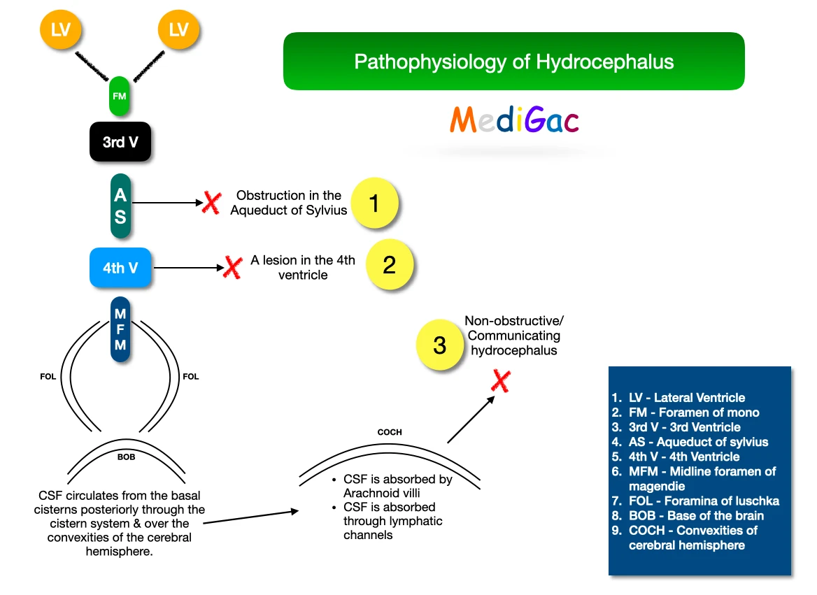
CSF flows into the 3rd ventricle through the foramen of monro from the lateral ventricles. Then it crosses the Narrow Aqueduct of sylvius and pours into the 4th ventricle. Then it exits the 4th ventricle and goes via two paired Foramina of luschka and Midline foramen of Magendie. Finally it goes into the cisterns at the base of the brain. Then CSF circulates from the basal cisterns posteriorly through the cistern system & over the convexities of the cerebral hemisphere.
- Obstruction in the Aqueduct of sylvius – Blocks the passage of CSF.
- Any lesion in the 4th ventricle also hampers the CSF passage.
- If any abnormality is occurring in the Convexities of cerebral hemisphere, then CSF absorption can get hampered.
4. Management :
A. Investigations :
- Taking history of family, which suggests X-linked or autosomal hydrocephalus secondary to aqueduct stenosis.
- Cranial bruit is audible.
- Plain Xray shows separation of sutures, erosion of the posterior clinoids.
- Ultrasound
- CT scan
- MRI
B. Treatment :
- Acetazolamide and Furosemide which reduces rate of CSF production.
- Endoscopic third ventriculostomy(ETV).
- Extracranial shunts.