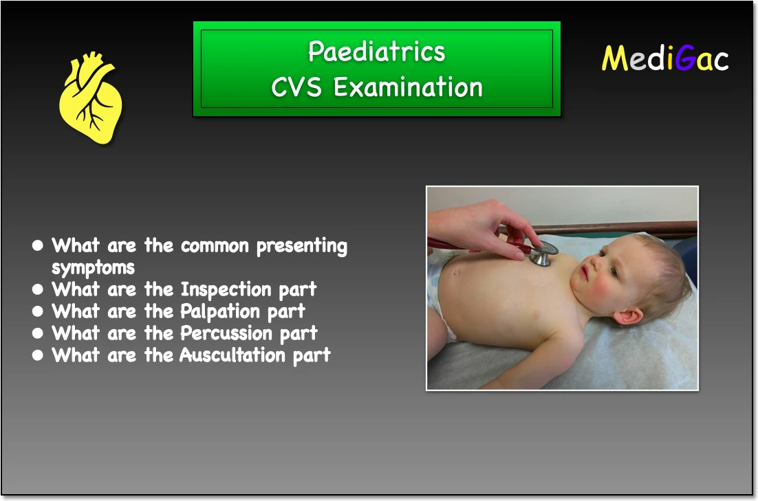
Paediatric cvs that is cardiovascular system examination. Examination of heart diseases by Inspection, Palpation and Auscultation. We are going to discuss four main cardiovascular system examination topics in paediatrics clinical examination.
- What are the common presenting symptoms
- What is the Inspection part
- What is the Palpation part
- What is the Auscultation part
I. What are the common presenting symptoms
1. If the patient presents with fever :
A newborn with fever, then he/she may be suffering from infective endocarditis, and pericarditis.
And it occurs like a swinging fever in paravalvar abscess.
2. If the patient presents with Sore throat :
Infections, such as a cold or flu, often cause pain or irritation in the throat, which can occur with or without swallowing.
Sore throat simply suggests Rheumatic fever carditis.
3. If the patient presents with Jaundice :
In newborn newborns, jaundice is a very frequent ailment. It frequently shows up in the first few days or weeks of a child’s existence.
If a newborn/child is suffering from jaundice, then this can be due to chronic congestive heart failure.
4. If the patient presents with pallor :
Skin or mucous membranes that are pale or have lost their colour.
Pallor is the occurrence of paleness of skin, this mainly occurs due to anaemic heart failure, and chronic heart disease.
5. If the patient presents with clubbing :
Changes in the areas under and around the toenails and fingernails that occur as a result of certain illnesses are known as clubbing. Changes in the nails are also seen.
Clubbing of fingers seen in congenital cyanotic heart disease(CHD)
6. If the patient presents with cyanosis :
Cyanosis is the bluish discolouration of the body, this can be central or peripheral.
- Central cyanosis – This mainly occurs in congenital CHD, and severe heart failure.
- Peripheral cyanosis – This can occur due to heart failure, cold and vasospasm.
7. If the patient presents with Lymph node enlargement :
Enlarged lymph node mainly occurs due to infection, and there can be various causes :
- Tubercular
- Bacterial infection
- Viral pericarditis
- Pericardial effusion
8. If the patient presents with Koilonychia (Spoon nails) :
- Spoon nails (koilonychia) are delicate, scooped-out nails. A drop of liquid may usually be held in the depression.
- Spoon nails are frequently a sign of iron deficiency anaemia or hemochromatosis, a liver ailment in which your body absorbs too much iron from the food you eat.
- This is mainly a nutritional problem.
9. Tender hepatomegaly :
- This occurs in congestive cardiac failure.
- The gap between the upper and lower border of the liver span is 12-15 cm in elderly children and 8-11 cm in children aged 4 to 8. From birth to three years, the neonate’s height is 5.5 – 7 cm.
10. Others :
- Roths spot : This can be observed by fundoscopy.
- Osler’s node : These are tender erythematous nodules on the pulp of fingers.
- Janeway lesion : These are painless erythematous lesions and present at the hypothenar area of palms.
- Oedema : This can be pedal, sacral and occurs in congestive cardiac failure.
- Base of the lung : These are fine crackles which occurs in left ventricular failure.
- Bed side proteinuria : This can occur due to glomerulonephritis in infective endocarditis.
- Cold extremities : This mainly occurs in congestive cardiac failure due to reflex vasoconstriction.
- Skin survey : Rash in Libbman’s sac endocarditis
- Rheumatic chorea
- Malar flush in SLE
III. What are the part of Inspection
In the inspection we check few things, and they are :
- Scars – Scars can be made due to any previous surgery like heart surgery, sternotomy, following mitral valvotomy, aortic valve replacement, infraclavicular scar after pacemaker insertion, and any left submammary scar are all visible.
- Impulse – Apical impulse(Apex beat) and Visible impulse or visible pulsation( Pulses coming from the heart or great vessels that are visible or felt on the anterior chest wall are known as precordial impulses.)
- Any dyspnoea or distress
- Dilated vessels
- Pectus excavatum(funnel chest) and Pectus carinatum(Pigeon chest)
- In coarctation of aorta we can see collateral arteries
- Harrison’s sulcus can also be observed as a big left-to-right shunt.
III. What are the part of Palpation
Palpation refers to the touching of patients with various areas of the hands, including gentle(This can be done by depressing the skin 1-2cm) and deep palpation(This is depressing the skin 4-5 cm with firm, deep pressure).
- Apical mitral impulse – Under the age of 5, this impulse is shown in the left 4th intercostal space, lateral to the midclavicular line, and after 5 years, it is visible in the 5th intercostal space, medial to the midclavicular line.
If there is left ventricular dilation, such as in severe hypertension, aortic stenosis, anaemia, or dilated cardiomyopathy, it might be moved inferiorly and laterally.
- Hypertrophic cardiomyopathy – The apical impulse is given a double surge by palpable 3rd and 4th cardiac sounds. Hypertrophic cardiomyopathy is characterised by a double apical impulse.
- Tapping apex beat – It is usual to see tapping apex in mitral stenosis and thrusting apex in aortic valve disease. However, this useful item has lost its value in recent years.
- Thrill of aortic stenosis – Aortic stenosis can be felt in the aortic area (right 2nd intercostal space) and the neoaortic area (third left intercostal space), as well as the apex and lower sternal edge or neck.
- Thrill of pulmonary stenosis – A pulmonary stenosis or patent ductus arteriosus (PDA) throb may be felt in the left 2nd intercostal region.
- Right ventricular heave – Right ventricular heave over the left parasternal edge is a sign of right ventricular hypertrophy(RVH) or dilation, which is frequently associated with pulmonary hypertension. Epigastric pulsation can be caused by RVH.
- Left parasternal heave – A systolic rise of the left costal cartilages caused by contraction of a hypertrophied right ventricle is known as left parasternal heave.
- Left ventricular and displacement of apex – Left ventricular dilatation (LVH) and apex displacement are caused by chronic volume overloading of the left ventricle due to mitral regurgitation and aortic regurgitation.
- Left ventricular aneurysm – This can be palpated at medial to cardiac apex sometimes.
IV. What are the part of Auscultation
Auscultation simply means listening with Stethoscope.
I. Heart sound :
Normally there are four heart sounds, S1, S2, S3 and S4
- S1 (‘lub’) & S2 (‘dub’) : The examiner begins by identifying S1 and S2 in each site. S1 is normally heard near the apex and S2 at the left sternal margin.
If there is any splitting of S1 and S2, it should be highlighted. Splitting is usually easier to hear in children than in adults.
- S3 (‘Tub-dub-dum’) :
- With the bell at the apex, you can hear a low-pitched early diastolic sound. It coincides with fast ventricular filling right after the A-V valves open, and is thus heard as Tub-dub-dum after S2.
- Pregenacy, anaemia, fever, and thyroroxicosis are all common findings in children and young adults.
- S4 (‘daa-lub-dupp’) :
Due to rapid ventricular filling by atrial contraction, the fourth sound is similarly a low frequency but late diastolic murmur.
S4 is a pathological condition that arises when the atrium contracts vigorously late in diastole to help fill the hypertrophied, noncompliant ventricle. Hypertension, aortic stenosis, and hypertrophic cardiomyopathy are the most common causes.
S4 is only physiological in a few circumstances.
II.Cardiac murmurs :
The sound of blood rushing through the heart, which can be caused by anything from regular heart exercise to a damaged heart valve or other abnormalities.
The diaphragm of a stethoscope can pick up high-frequency sounds like aortic regurgitation, whereas the bell of a stethoscope can pick up low-frequency sounds like mitral noises.
This is heard over the right second intercostal space that is the aortic area na dover the left sternal edge that is the neoaortic area.
- Mid diastolic murmur – This happens when there is significant aortic regurgitation and the regurgitant jet closes the mitral valve. This is best heard with bell of the stethoscope. In VSD and MR, increased blood flow through a non-stenotic mitral valve causes a mid-diastolic murmur.
- Innocent Murmur – These are mid-systolic systolic systolic systolic systolic systolic systolic systolic systolic systolic systolic systolic systolic systolic systolic systolic systolics induced by turbulent flow tracts, usually
- Pansystolic murmur – Mitral regurgitation, tricuspid regurgitation, and ventricular septal defects are the most prevalent causes of pansystolic murmur, which is a regurgitant murmur heard throughout systole due to blood flow between two chambers with generally quite different pressures in systole.
- Continuous murmurs – In patent ductus arterisus at the base of the heart, they can be heard in both systole and diastole. And a valsalva aneurysm with a burst sinus. This is most noticeable at the end of systole and gradually fades throughout diastole.
- VSD, TR murmurs are heard loudest at the left lower sternal edge.
III. Others :
- At apex – Mitral regurgitation
- Left lower sternal border – tricuspid area
- Pulmonary area – we can listen for ASD, pulmonary stenosis and pulmonary regurgitation.
- Aortic area – We can listen for aortic stenosis, hypertropic cardiomyopathy, and regurgitation.
- Tricuspid area – Tricuspid regurgitation and tricuspid stenosis.