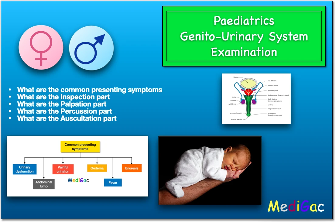
Genito urinary system examination of paediatrics or neonatology. Nephrology of paediatrics. common presenting symptoms, inspection, palpation, percussion and auscultation.
We are going to discuss the topics related with genito urinary system examination. We have discusses five topics and they are described below.
- What are the common presenting symptoms
- What are the Inspection part
- What are the Palpation part
- What are the Percussion part
- What are the Auscultation part
I. What are the common presenting symptoms :
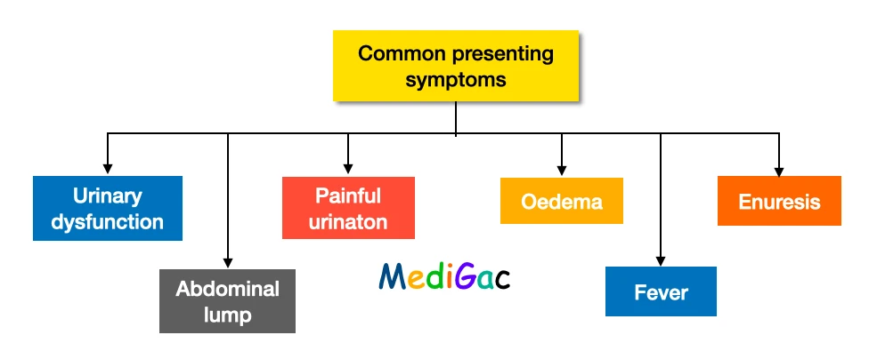
There are some common presenting symptoms of genital and urinary system diseases or disorders. They are described below in details. We have discussed total of six common presenting symptoms.
I. Urination dysfunctions :
Urinary dysfunction can occur in three various properties of urine.
1. Depending upon urine volume :
Urinary volume dysfunction can be of two types – Oliguria and Polyuria
- Oliguria – When urine volume is less than normal. This can occur due to Acute glomerulonephritis, Childhood nephrotic syndrome, and Haemolytic uraemia syndrome.
- Polyuria – When urine volume is more than normal value and approximately about more than 4ml/kg/hour. This can occur due to chronic renal failure, renal tubular acidosis, central diabetes insipidus, insulin-dependent diabetes.
2. Related with micturition :
With the characteristics of micturition, we can divide the dysfunctions into five categories, and they are – Dysuria, Urine retention, urination with poor stream, sudden micturition, increased frequency of micturition, and Persistent dribbling of micturation.
- Dysuria – This is when the patient cries or shows agony while micturition. The causes can be urinary tract infection, bladder outflow gets obstructed due to posterior urethral valve and bladder neck obstruction.
- Urine retention – Urinary retention is a condition in which a person’s bladder is unable to empty completely. And this can occur due to UTI, posterior urethral valve, and neurogenic bladder( lack of bladder control due to nervous system disorder)
- Urination with poor stream – In posterior urethral valve in males can run urine passing drop by drop.
- Sudden micturition – When the urge to micturition occurs, the patient may not be able to contain the urine. UTI, bladder stone, and vaginitis in girls are the most common causes.
- Increased frequency of micturition – Urine output does not surpass the normal limit after 24 hours, despite increased frequency of passing urine. The causes are UTI, and bladder stone.
- Consistent dribbling – continuous dribbling of urine, due to abnormal urine drainage to urethra and neurogenic cause.
3. Depending upon the colour of the urine :
Most common colour of dysfunction urine is Red. And the causes are – Haematuria, Myoglobinuria, Pigmenturia and Haemoglobinuria.
Behind haematuria, there are several causes like tumour, nephrolithiasis, Cystitis, Glomerulonephritis.
II. Abdominal lump
A lump in the abdomen is a swelling or protrusion that appears in any part of the abdomen. The bulge is visible in the lumbar region and can be manually palpated and balloted. Hydronephrosis, Wilms tumour, Renal vein thrombosis, Polycystic kidney disease, and Multicystic renal disease with renal dysplasia are some of the causes.
III. Painful urination
There are mainly two types of pain and they are – Pain over loin, hypogastrium and Colicky pain.
- Pain over loin and hypogastrium – The loin pain mostly occur in UTI mostly and it dull aching type of pain. And the hypogastrium pain occurs due to cystitis.
- Colicky pain – Colicky pain in adults is typically a strong, localised urine discomfort that might start suddenly and comes and goes in spasmlike waves. This pain starts at loin and then spreads towards groin and also then to thigh and genitalia. The causes are rena; and ureteric colic which usually occurs due to kidney stone.
IV. Oedema
Oedema that is fluid filled swelling first occurs at the face, known as facial puffiness and then starts to appear over legs and feet. Finally it spreads all over the body.
The causes of oedema are – Acute glomerulonephritis, Acute and chronic renal failure, and Nephrotic syndrome.
V. Fever
This is a common feature of infection, so in UTI this occur so frequently. There are two specific disease in which the fever varies, and they are – Pyelonephritis and Cystitis.
- Pyelonephritis – here the fever appears high and commonly associated with chill and rigor.
- Cystitis – There may or may not be a fever present.
VI. Enuresis
Enuresis is the medical term for not being able to regulate your pee. Enuresis is also known as involuntary urination. Involuntary urine that occurs at night while sleeping, after a person has reached the age when he or she should be able to regulate his or her bladder, is known as nocturnal enuresis.
There may be presence of primary and secondary enuresis.
- Primary enuresis – When a child has never been on a continent, this can happen. This is mainly physiological and occurs mostly as nocturnal.
- Secondary enuresis – When enuresis appears after a child has been continent for at least 6 months. Common causes are – UTI, spina bifida, diabetes mellitus, posterior urethral valve. This are responsible for almost 20% of cases.
II. What are the Inspection part :
1. Scars :
This is seen in kidney transplants, ambulatory peritoneal dialysis nephrectomy, and intermittent and continuous peritoneal dialysis nephrectomy.
2. Abdomen :
- Ascites, a mass such as an enlarged kidney or bladder, causes a distended belly with fluid.
- In the right iliac fossa, the transplanted kidney may be visible.
- Abscess, hydronephrosis, and polycystic kidney disease can all cause fullness in the loin.
3. Bladder :
- In acute retention and obstructive uropathy, a distended bladder is seen.
4. Kidney :
- Obstructive uropathy, hydronephrosis, polycystic kidney disease, Wilm’s tumour, and renal vein thrombosis all cause increased kidney size.
- The transplanted kidney may be visible in the right iliac fossa.
5. Genital:
- Balanitis – This is inflammation of the glans penis, is a frequent illness that affects between 3 and 11 percent of men.
- Balanoprosthitis – Balanoposthitis, defined as inflammation of the foreskin and glans in uncircumcised males, affects people of all ages and can be caused by a variety of bacteria, fungi, or contact dermatitides.
- Valvitis – Vulvitis is a condition in which the vulva becomes inflamed. Outside the vagina, these are the delicate folds of skin. It’s a symptom that might appear as a result of a variety of illnesses.
- Scrotal or labial edema – This oedema happens in people with nephrotic syndrome, thus it’s important to protect the privacy of an older child.
- Phimosis, Pinhole meatus
- Hypospadias and Epispadias
III. What are the Palpation part :
During a physical examination, palpation is a means of feeling with the fingers or hands. The health care professional examines the size, consistency, texture, location, and tenderness of an organ or body part by touching and feeling it.
1. Kidney :
- Right kidney – The examiner places his or her left hand in the loin area (between the 12th rib and the ileum bone) and crosses his or her left hand over the anterior abdominal wall. The examiner next instructs the child to take a deep breath and feel for the lower pole of the kidney going down between the palms. The examiner then uses his or her hands to gently push the kidney back and forth to demonstrate movement. Ballotment is the term for this procedure.
- The process is repeated to ballots the left kidney and left renal angle.
- We used to palpate the liver incase of PKD(Polycystic kidney disease).
- In the right iliac fossa, the transplanted kidney can be palpated.
- A polycystic kidney enlarges in the front, but a perinephric abscess enlarges in the rear.
2. Renal angle :
The renal angle is gently and firmly palpated between the spine and the 12th rib on the posterior side. If there is no discomfort, the examiner strikes the renal angle firmly with moderate force with the ulnar aspect of the fist. It’s called Murphy’s Kidney Punch, and it’s not too hard.
3. Bladder :
- The examiner palpates the upper, right, and left borders of the urinary bladder and delinates them.
- The use of two hands to palpate the bladder is more reliable.
IV. What are the Percussion part :
Percussion is a physical examination technique that involves tapping bodily areas with fingers, hands, or tiny instruments. It is carried out in order to ascertain:
- Body organs’ size, consistency, and borders
- The presence or absence of fluid in various parts of the body.
- On the midline – A percussion in the midline is used to see the upper boundary of the bladder, which aids in determining whether it is distended or full.
- We need to look for ascites-related shifting dullness.
V. What are the Auscultation part :
Listening to noises from the heart, lungs, or other organs with a stethoscope as part of a medical diagnosis.
- Renal bruits – These are heard in the transpyloric plane, which is lateral to the rectus abdominis, and can be difficult to distinguish from abdominal vascular bruits. The inspector also tries to listen to it from the back of both loins. There are bruits in 50% of renal artery stenosis. The diastolic bruit is more suggestive in this case.
- It’s also a good idea to listen to the sound of your bowels.