Paediatrics, neonatology respiratory system examination. Inspection-Palpation-Percussion-Auscultation. And the common respiratory problems.
We are going to discuss five main topics related with respiratory system examination.
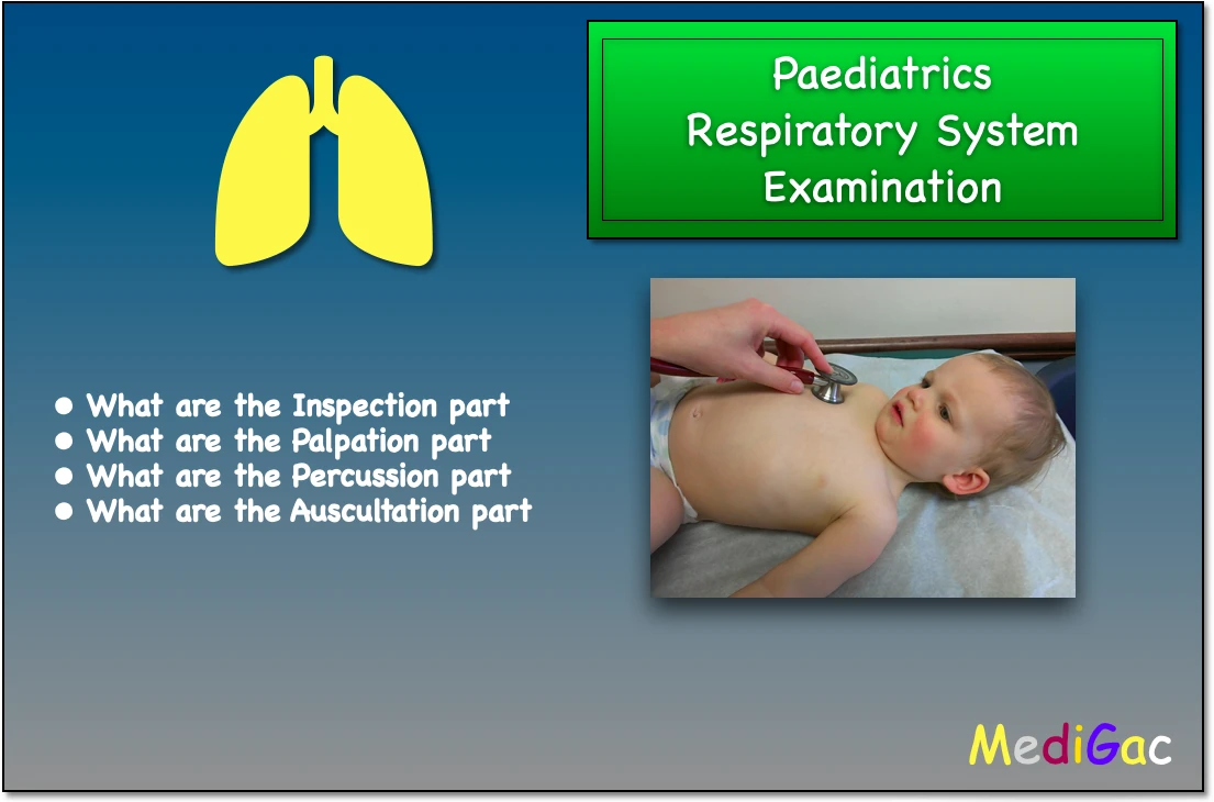
- What are the Inspection parts
- What are the Palpation parts
- What are the Percussion parts
- What are the Auscultation parts
I. What are the Inspection parts
In inspection part we mainly focus on four things or topics, that is scars, any signs, Respiration and the chest.
I. Scars :
- Any previous surgery can make scars, which are seen in present. Surgeries like Lobectomy, Tracheostomy, Cardiac surgery like Thoracotomy, Pulmonary artery banding, BT shunt .
- Biopsy mark at the level of axilla
- Water seal drainage scar which was used in pneumothorax
- Any scar of drainage tube which was given in empyema thoracic, and huge pleural effusion
II. Any specific signs :
- Pemberston’s sign – The tester instructs the child to raise his arms over his head and then wait one minute. Then there’s facial plethora, cyanosis, inspiratory striador, and high JVP in the event of SVCO (Superior venacava obstruction).
- Kyphosis – Exaggerated anterior curvature of the spine is seen in tuberculosis and poliomyelitis of the spine.
- Kyphoscoliosis
III. Respiration :
- First we have to check the respiratory rate, whether it is Normal/Rapid or Shallow.
- Secondly we will check for the types of respiration, whether it is Abdomina, Abdominothoracic, Thoracic, or Thoracoabdominal.
IV. Chest :
- Pectum excavatus and Pectus carinatum
- Symmetry or asymmetry of chest
- Shape of the chest – Barrel shaped chest (The AP diameter > lateral diameter, and occurs in hyperinflation of lungs like in COPD, Recurrent acute severe asthma ).
- Chest movement – If the chest movement gets restricted, this occurs in Pleurisy and Pneumonia.
- Flattening of chest is seen
- Skin of chest should be carefully examined
V. Others :
- Bulging of intercostal space is seen in pleural effusion.
- Subcostal recession – IMCI suggests subcostal recession as a sign of pneumonia.
- Rhythm should be checked, whether it is Regular, Irregular or others like Chynestoke
II. What are the Palpation parts
Palpation is done in three line that is Midclavicular line, Midaxillary and sub scapular line.
- Lymph nodes – The lymph nodes at chest and axillary to be palpated carefully.
- Tenderpoint in the chest wall – This occurs in rib fracture, any inflammatory or malignant condition, costochondritis of Tietze syndrome or any infective condition.
- Any swelling should be palpated
- Chest expansibility should be palpated by placing the hands, extended fingers and thumb.
- Vocal fremitus – A vibration conveyed through the body is known as vocal fremitus. It refers to evaluating the lungs by feeling the intensity of vibrations on the chest wall (tactile fremitus) and/or hearing particular uttered words through a stethoscope on the chest wall (vocal resonance).
- Tracheal tug – During inspiration, tracheal tugging is defined as an aberrant downward movement of the trachea accompanied by in-drawing toward the thoracic cavity.
- Paradoxical inward movement may suggests diaphragmatic paralysis and severe COPD.
- Tracheal and cardiac apex shift – This indicates a mediastinal movement, while the index finger detects a tracheal shift. Along with this, the apex beat should also be felt.
III. What are the Percussion part
To make percussion in the mediclavicular, midaxillary, and posterior scapular lines, the examiner uses a swinging movement of the wrist to push his or her middle finger perpendicularly against the tip of the right hand’s middle finger.
Then the sound, resonance, pain, and if any tenderness is present should be noted.
- Right side liver dullness – This interferes and left sided stomach gas makes more resonance.
- Then the examiner compares both sides at leats 3-4 sites on 3 lines, that is mid-clavicular, mid-axillary and posterior scapular.
IV. What are the Auscultation parts
In auscultation part, we mainly focus on the breath sounds. To hear the breath sounds, we use the diaphragm part of the paediatrics stethoscope.
Above the clavicle, along the mid clavicular line up to the 6th rib, laterally along the mid axillary line up to the 8th rib, and posteriorly up to the 11th rib, the breath sound can be heard. It is advised to auscultate the both side breath sounds and comparing them simultaneously.
There are mainly three types of breath sounds –
- Bronchial breath sound – This sound is heard on consolidation more louder and harsh.
- Vesicular breath sound – This is normal and occurs like 3 seconds in inspiration and 2 seconds in expiration.
- Vesicular breath sound – Vesicular with prolonged expiration like in asthma and COPD.
There are presence of four additional or specific sounds which are pathological also.
- Wheezing
- Crackling sound
- Stridor
- Pleural rib
I. Wheezing :
This can occur due to asthma and COPD.
Monophonic wheezing – It is caused by the constriction of a single bronchus due to foreign bodies and tumours. It can be inspiratory, expiratory, or both, and its intensity can alter according on the position.
II. Crackling sounds :
Short, explosive sounds that are often described as bubbling or clicking. Even without a stethoscope, a coarse rattling sound can be heard when big airways are clogged with sputum.
- Fine crackles – These are found in left ventricular, pulmonary edema, and diffuse interstitial fibrosis.
- Coarse crackles – This suggests pneumonia, and bronchiectasis.
III. Stridor :
This could be a sign of laryngitis or a constriction of the trachea or big airway. This can be heard without a stethoscope and is both inspiratory and expiratory. It usually signals a serious underlying ailment.
IV. Pleural rub :
On auscultation of the lung, a pleural friction rub is an unexpected breath sound. The sound of pleural rub is caused by the movement of inflamed and roughened pleural surfaces against one another while the chest wall moves.
This is heard in :
- Pneumonia
- Pulmonary vasculitis
- Pleural effusion
V. Other sounds :
- In severe airway obstruction, a silent chest is noted.
- When there is air between the two layers of pleura overlaying the heart, a pneumothorax is a rhythmical sound synchronised with cardiac systole.
- As the patient repeats nine times, the stethoscope detects sound vibrations delivered to the chest from the voice cords.
- If both the pleura and the pericardium are inflamed at the same time, pleuropericardial friction rub is audible.