We have made all the RADIOLOGY Instruments full set or list with the Names, Description, Uses with Pictures. in you medical ward you will see all this instruments/equipments/devices. So full knowledge regarding all this devices is necessary for your medical and surgical practice and also during exams.
Nuclear Magnetic Resonance
Description :
This machine works by combination of different components :
- Computer
- Spectrometer
- Super-Conducting Magnet
Uses :
Advanced magnetic resonance imaging.
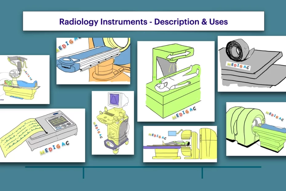
Brachytherapy Apparatus

Description :
This device is made for sending radioactive source in the body.
—The radioactive source travels from brachytherapy machine through hollow tubes or needles
Different parts :
- Motor
- Fixing Rod
- Lead Screw
- Coupler
- Needle Template
- Needle
Uses :
1. Internal Radiation Therapy : To treat cancers of head and neck, breast, prostate and eye.
Computer Axial Tomography Machine

Description :
This machine helps to take detailed radiological pictures of the body/internal organs.
Uses :
1. Diagnosis of diseases
2. Planning treatment
3. Finding out how well the treatment is working
Functional Magnetic Resonance Imaging

Description :
This device works by fMRI using the Bool-Oxygen-Level dependent contrast.
Uses :
Measures brain activity by detecting change associated with blood flow.
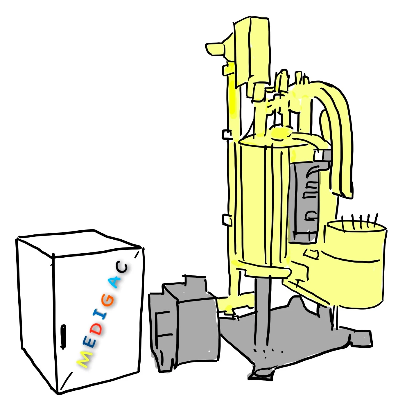
Lead Shielding
Description :
Made up of lead material and used for shielding Walls, Floors, Radioactive machines.
Uses :
Give protection against radiation by high shielding property.
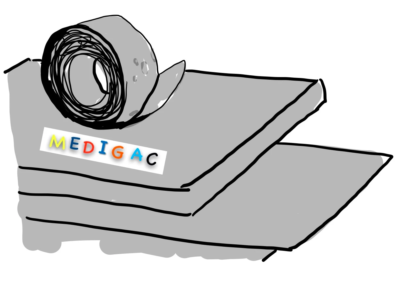
Linear Accelerator
Description :
This device is acts as a particle accelerator which helps to accelerate charged ions in high speed.
Uses :
1. Treatment : Delivers external beam radiation to treat cancer.
Magnetic Resonance Imaging

Description :
This device uses strong magnetic fields along with strong radio waves.
—–This combination helps to get detailed images of the inside of the body.
Uses :
To see detailed structures of any part of the body.
Positron Emission Tomography

Description :
This device is a functional imaging technique which uses radiological substances to observe and measure :
- Physiological activities like metabolism processes, Blood flow, etc.
Uses :
Using a radioactive drugs this device helps revealing how the issue and organs are functioning.
Radio Isotope Scan

Description :
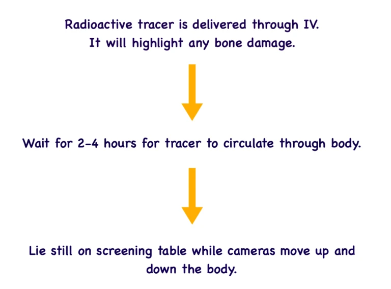
- Radioactive tracer is delivered through IV. It will highlight any bone damage.
- Wait for 2-4 hours for tracer to circulate through body.
- Liestill on screening table while cameras moves up and down the body.
Uses :
Used small dose of radioisotope to detect cancer, trauma, infection or other disorders.
SPECT Scan

Description :
SPECT : Single-Photon emission computerised tomography scan helps to see the doctor inside the body.
Uses :
- Brain Disorders
- Heart Problems
- Bone Disorders
Ultrasonography(USG) Machine

Description :
Used mainly during the Gestation period. Has different components :
- Monitor
- Onboard Computer
- Pulse controls
- Keyboard
- Transducer
Uses :
Used to see inside the Pelvis or Abdomen mainly:
1. In Pregnancy or during ,12 Weeks of Gestation.
2. Any kind of GI problems, sometimes we do USG.
X-Ray Machine

Description :
This machine works by generating X-rays.
Uses :
In Diagnosis of :
- Bone Disorders : Bone fractures, Arthritis, Bone Tumours
- Infections such as Pneumonia
- Calcifications like Kidney Stones
Interventional Radiology

Description :
This holds all the radiological imaging procedures performed for doing any kind of treatment.
—- Instead of giving incision and putting scope, the doctors see inside the body by CT, MRI,USG, and fluoroscopy during any treatment procedure.
- X-Ray Fluoroscopy
- Computer Tomography
- Magnetic Resonance Imaging
- Ultrasonography
Uses :
1. Angioplasty : Repairing or unblocking of blood vessels
2. Stenting : Small mesh tubes that treat narrow or weak arteries.
3. Thrombolysis : Dissolving blood clots
4. Embolization : Block blood flow to cancer cells
5. Biopsy : Studies of tissues.
Mammogram

Description :
This is a large device and has mainly 3 parts :
- Camera Unit
- X-Ray Beam
- Film Plate
Uses :
This is mainly used for detecting beast abnormality likes breast cancer, palpable masses and nonpalpable breast lesions.
Instruments’ Page