Know how to examine the peripheral arterial including Present Complaints, General and Physical examinations, in the cardiovascular system.
Examination of the Peripheral Arterial System
1. What are the symptoms, a patient can present with ?
The patients with peripheral vascular system disorders can present with the, Abdominal pain, Leg pain, Digital ischaemia, Stroke.
I. Pain in the abdomen :
- Aortic Aneurysm – At the level of the abdomen, an expansion of the aorta, the primary blood vessel that carries blood to the body.
- Arterial blockage – A blockage in an artery causes mesenteric ischemia, which prevents blood flow to a part of the intestine.
II. Feeling pain in the leg :
- Pain during sleeping at night – After sleeping 1-2 hours, the patient again wakes up after starting to feel the severe pain in foot, usually in the instep.
- Silent myocardial ischaemia – When atherosclerosis affects the large and medium sized blood vessels, then this kind of pain can occur. Most of the time remains asymptomatic.
- Building of pressure – Internal bleeding or tissue swelling causes pressure build-up, which is uncomfortable and dangerous. This is also known as compartment syndrome.
- Exercise time – When the arteries are restricted or clogged, blood flow to the legs is reduced. Claudication pain appears when the person walks a particular distance or does some exercise and then it disappears when he/she starts to take rest.
- Pain during relaxing time -This kind of pains can the blood flow can’t able to meet the demand of the tissues.
- Decrease in limb perfusion – This is the sudden decrease in the perfusion.
- Abdominal Aortic Aneurism – At the level of the abdomen, an expansion of the aorta, the primary blood vessel that carries blood to the body.
- loss of continuation of tissues or Ulcer– Perfusion is insufficient to maintain the tissues in individuals with severe lower limb PAD, and tissue death (gangrene) develops at the tips of the digits, gradually migrating proximally.
III. Ischaemia disfigurement :
- Blue toes – When a AAA(Abdominal Aortic Aneurysm) or another proximal embolic source, such as a popliteal aneurysm or atherosclerotic plaque, causes atheroembolism, blue toe syndrome develops.
- Vasospastic Symptoms – Raynaud’s syndrome is a type of digital ischaemia brought on by cold and emotion. There are three stages to it, they are pallor, cyanosis and redness.
Pallor – It occurs by obstruction of the digital artery
Cyanosis – Occurs when body fails to oxygenate the venous static blood.
Redness – Occur due to relative hyperaemia(Increased blood flow to a part of the body.)
IV. If patient presents with Stroke :
Interruption of the brain’s blood flow causes damage, which can end by resulting Stroke.
Stroke symptoms include difficulty walking, speaking, and understanding, as well as facial paralysis or numbness.
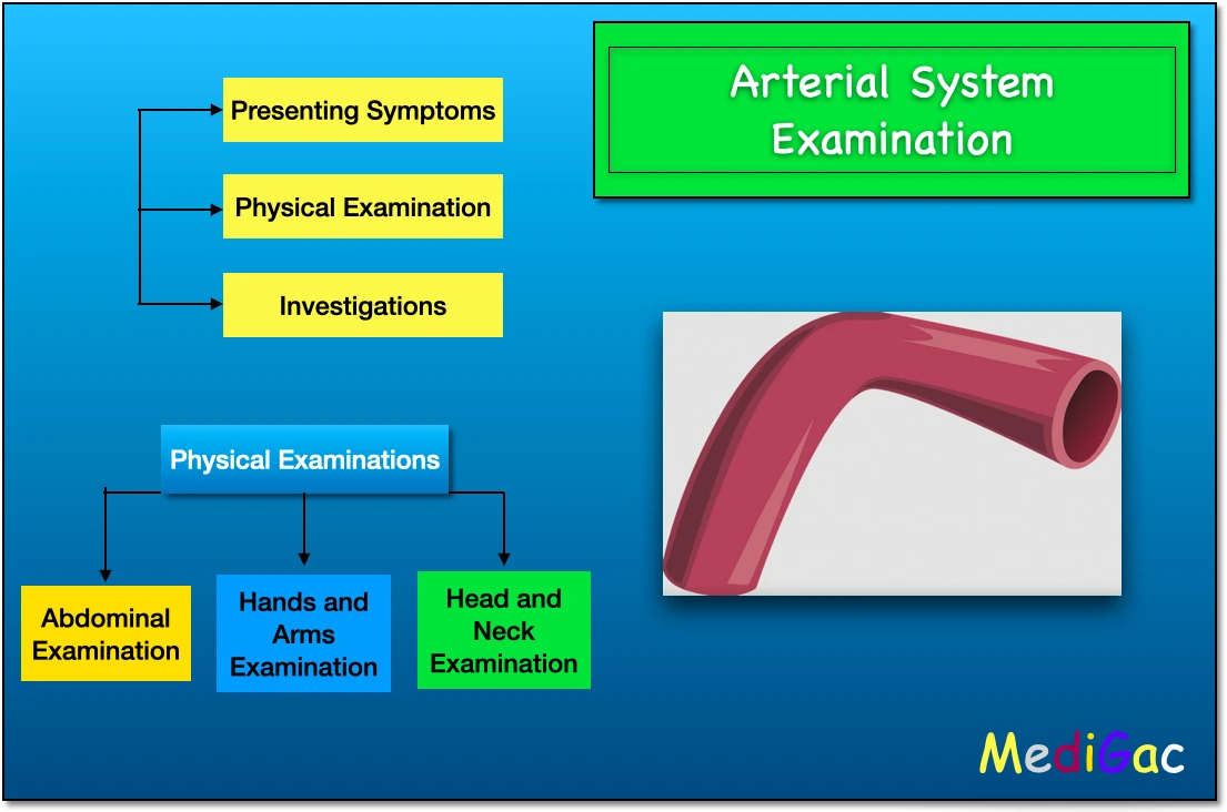
How to examine such patients physically :
So for the physical examination of this patients, we must have to see the signs in three sites, Abdomen, Hands and Arms, & Face and Neck.
The we have to follow a Examination sequence that is :
Arm(Radial & Brachial Pulse with Blood Pressure measurement) –> Neck(Carotid Pulse) –> Abdomen(Palpating the Abdominal Aorta, Aortic bifurcation of Umbilicus) –> Legs(If any scars or ulcers are present) –> Femoral Pulse –> Popliteal Pulse –> Posterior Tibial Pulse –> Dorsal Tibial Artery Pulse
I. Examination of the Abdomen :
- Epigastric or Umbilical Pulsation – Here you just have to palpate the abdominal aorta, and it suggests ‘Aortoilliac Aneurysm‘
- Weight loss – The patient comes with complaining weight loss, then it can suggest Visceral Ischaemia
- Mottling of the abdomen – splotches or patches of various tints or colours, signifies Aortic Aneurysm Ruptured abdominal aortic aneurysm.
II. Examination of the Hand and Arms :
- Finger Tips – If purple discolouration is present, then it suggest the Atheroembolism from proximal subclavian aneurysm
- Finger Pulps – If there are presence of Pits and healed scars then it suggest Secondary Raynaud’s Syndrome(In response to cold temperatures or stress, the fingers, toes, ears, and tip of the nose are typically affected and feel numb and chilled)
- Tobacco Stains – When brown tobacco is combined with saliva, it produces a dark brown liquid that stains easily. The staining power of such a liquid will leave a lasting impression on the teeth with which it comes into contact.
- Small muscles of Hand wasting – This can signifies Thoracic outlet syndrome.
- Capillary Loops – This suggests, Calcinosis, Oesophageal dysfunction, Telangiectasia, Raynaud’s phenomenon.
III. Examination of the Face and Neck :
- Corneal arcus and Xanthelasma – This suggests Hypercholesterolaemia
- Hoarseness of the voice along with ‘bovine’ cough(a non-explosive cough caused by an inability to seal the glottis) – This suggests Recurrent laryngeal nerve pals from a thoracic aortic aneurysm.
- Prominent veins in the neck, shoulder and anterior chest – This suggests Occlusion of the Axillary/Subclavian vein
- Horner’s Syndrome – This is occurs when on one side, the neural route from the brain to the face and eye is interrupted. And it signifies the Carotid artery aneurysm.
Two special tests are done :
1. Buerger’s Test :
The Buerger’s test is performed to evaluate arterial sufficiency. When lying down, this is the angle at which the leg must be elevated before it appears pale. Even when the limb is lifted 90 degrees, the toes and sole of the foot remain pink in a limb with proper circulation.
2. Ankle Brachial pressure index :
The ankle-brachial index (ABI) is the ratio of blood pressure in the ankle to upper arm blood pressure (brachium). Lower blood pressure in the leg compared to the arm indicates clogged arteries owing to peripheral arterial disease (PAD).
What are the Investigations to be done :
For unilateral disease, duplex ultrasound is frequently the initial line of examination, whereas a CT or MR angiography might be used to evaluate bilateral symptoms.
- Duplex ultrasound – Carotid artery stenosis, abdominal aortic aneurysm monitoring, and peripheral arterial disease are all conditions that need to be monitored.
- Magnetic resonance imaging– Arteriovenous malformations, carotid artery stenosis
- Computed tomography – Abdominal aortic aneurysm, carotid artery stenosis
- Angiography – Carotid artery stenosis, acute and chronic limb ischaemia As a diagnostic test, invasive angiography has mostly been supplanted by computed tomography/magnetic resonance angiography.