In a new born baby or a mature children pulse checking can bring some important data regarding various diseases. Pulse rate, Rhythm, volume, characters are discussed.
We mainly focus on seven sites to check the pulse of the body :
- Radial pulse – Either wrist can be used to take the radial pulse. The examiner feels the pulse in the patient’s radial artery between the wrist bone and the tendon on the thumb side of the patient’s wrist with the tip of the index and third fingers of the other hand.
- Brachial pulse – The brachial pulse can be felt in the area of the antecubital fossa by pressing on the bicep tendon.
- Femoral pulse – The best place to feel the femoral pulse is in the inner thigh, halfway between the pubic symphysis and the anterior superior iliac spine, at the mid-inguinal point.
- Popliteal pulse – Felt at popliteal fossa
- Carotid pulse – The carotid pulse (CP) is a pressure signal obtained as the carotid artery passes near the body’s surface at the neck.
- Dorsalis pedis arterial pulse – On the dorsal aspect of the foot, the dorsalis pedis artery pulse can be palpated lateral to the extensor hallucis longus tendon or medially to the extensor digitorum longus tendon.
- Posterior tibial pulse – The pulse of the posterior tibial artery can easily be felt halfway between the medial malleolus and the Achilles tendon.
During pulse checkin we have to check the points :
- Rate – Normal/bradycardia/tachycardia
- Rhythm – Regular or Irregular
- Volume – Normal/Low/high
- Character
- Radiofemoral/radioaradial delay
- Vessel wall’s condition
1. Pulse Rate :
This is counted as number of times the heart beats per minute. The top limit for newborns is 200 beats per minute, the lowest limit is 70 beats per minute, and the average is 120-140 beats per minute.
Depending upon high and low pulse rate, it is mainly divided into two types that is bradycardia and tachycardia.
A. Bradycardia :
This occurs when the heart rate falls down than the normal heart rate. Bradycardia can be of two types that is Sinus bradycardia and Other bradyarrhythmias.
Sinus Bradycardia
This occurs when a newborn’s heart rate goes below 90 beats per minute and falls below 60 following the neonatal period.
Causes :
- During sleeping of a newborn baby
- LBW babies
- Hypothyroidism
Other Bradyarrhythmias
This can be caused due to various reasons :
- Sick sinus syndrome
- Congenital heart block
- second degree heart block
B. Tachycardia :
This condition is known by the increase in heart rate than the normal range. This varies upon the age.
- >200/min : In newborns
- >150/min : In infants
- >120/min : In older children
Now, here also tachycardia can be two types – Sinus tachycardia and Other tachyarrhythmias.
Sinus tachycardia
In this condition the rate variation maintained with the inspiration and expiration.
Causes :
- Heart failure
- Fever, exercise, and excitement
- Myocarditis
- Other inflammatory causes like rheumatic fever, diphtheria.
Other tachyarrhythmias
This can occur due to various causes :
- Atrial fibrillation
- Atrial flutter
- Paroxysmal supraventricular tachycardia(PSVT)
2. Pulse character :

There are mainly six character of pulse : Water hammer pulse, Pulsus paradoxus, Pulsus alternans, Anacrotic pulse, Pulsus bisferiens, Pulsus bigeminus.
We are starting to discuss from below :
I. Water hammer pulse :
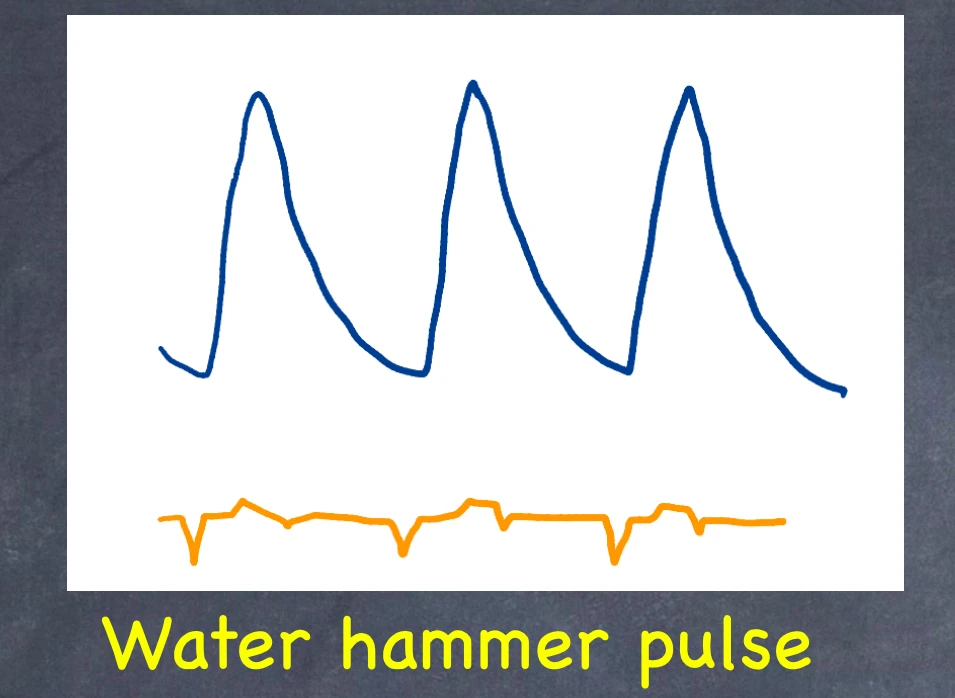
This is the corrigan pulse, which is the sharp rise and steep fall of the pulse wave.
II. Pulsus paradoxus :

This is a condition in which the pulse volume drops during inspiration and rises during expiration.
III. Pulsus alternans :

This indicates that in acute left ventricular failure, alternative pulse waves are weaker.
IV. Anacrotic pulse :
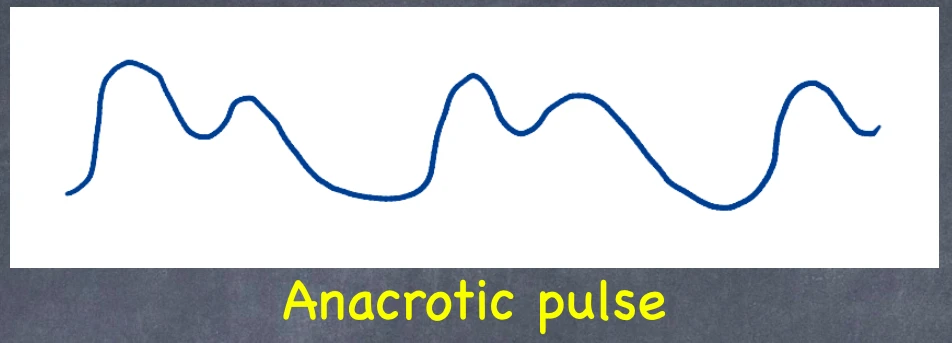
When the upward slope of the pulse wave has an anacrotic notch, such as in aortic stenosis, it is perceived as a slowly rising double beating pulse.
V. Pulsus bisferiens :

In hypertrophied obstructive cardiomyopathy, severe AR with mild AS, this is a double kicking or twice beating pulse during systole. The ideal place to feel this pulse is over the brachial artery.
VI. Pulsus bigeminus :
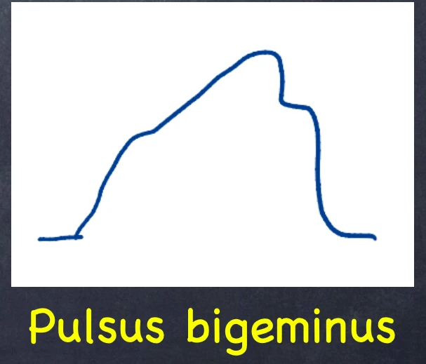
In heart block, there are two beats in a row followed by a compensatory halt in a regular cyclic pattern, such as 2:1. A powerful beat is followed by a weak beat in this arrangement. Pulsus alternans lacks this compensating pulse.
3. Pulse Rhythm :
This is the regularity of the pulse or the space between heart beats.
This can be of two types – Regular and Irregular rhythm
- Sinus arrhythmia – This is a physiological situation in which the heart rate slows during expiration and speeds up during inspiration. This is very frequent among children and young people.
- Pathological arrhythmia – Irregularly irregular pulse and regularly irregular pulse.
Pathological arrhythmia :
- Irregularly irregular pulse – This is due to atrial fibrillation, and multiple extrasystoles.
- Regularly irregular pulse – This occurs when in regular intervals irregularity is palpated. Like in heart block and atrial flutter.
4. Pulse Volume :
A blood pressure cuff and a hand-held Doppler ultrasound equipment are used in pulse volume recording (PVR) to detect the existence and severity of peripheral artery disease (PAD). The Doppler ultrasound measures the speed and flow of blood by recording sound waves that bounce off moving objects.
| High volume / Bounding pulse | Low volume or Thready pulse |
| 1. Aortic regurgitation 2. Arteriovenosu fistula 3. Aorta-pulmonary window 4. All hyperactive circulatory states like exercise | 1. Any cardiac lesions : Aortic stenosis, Heart failure,Obstructive cardiomyopathy, Constrictive pericarditis 2. Cardiac shock |
5. Radiofemoral-radioradial delay :
A. Radiofemoral delay :
The radial and femoral pulses are normally palpated at the same time. Radiofemoral delay is defined as a noticeable delay in the femoral pulse relative to the radial pulse.
B. Radioradial delay :
Situation is normal. The radial and femoral pulsations are felt in the same way and at the same time. Radio radial delay is the difference between two radial pulses. Radiofemoral delay is the time difference between the radial and femoral pulses.
6. Vessel’s Walls condition :
The state of the arterial wall
After digital pressure flattens an artery, it is no longer palpable. Arteriosclerosis is indicated by a thick radial artery perceptible 7.5–10 cm up the forearm.