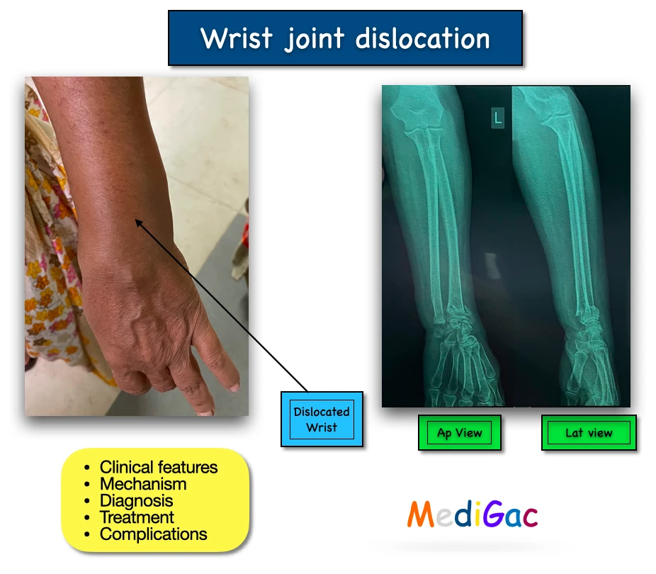
1. Clinical features :
- Swelling.
- Pain.
- Numbness and tingling.
- Deformity around the injured area.
2. Mechanism :
- Fall from height.
- RTA.
- Falling in hyperextended and pronated wrist.
Others associated conditions can be :
- Open fractures.
- Medial nerve injury > Ulnar nerve injury.
- Distal radius fractures.
- Ulnar styloid fracture.
- Carpal bone fracture.
- Intercarpal injury.
3. Diagnosis :
- Plain X-Ray of wrist – AP, lateral and oblique view.
- Physical appearance.
- CT scan.
- MRI is done to evaluate the integrity of the scapholunate and lunotriquetral.
4. Treatment :
There are mainly two types of treatment methods and they are – Nonoperative and Operative.
A. Non-Operative :
This is done by closed reduction or traction and providing caster slab for 5-6 weeks. This is mainly done when the patient is not medically stable for surgery and chance of getting stable radoiocarpal joint after traction.
B. Operative :
- Open reduction.
- Internal fixation.
- Radiocarpal pinning and ligament repair.
5. Complications :
- Malunion.
- Non union.
- Stiffness.
- Acute carpal tunnel syndrome.
- Post-traumatic arthritis.
- Chronic radiocarpal instability.
- Late inter carpal disruption.