Ulnar fracture is very common after falling form height or any accident. Ulnar fracture can be of various types based on the location and severity of the fracture. Here we have discussed the Proximal ulnar fracture.
1. Clinical features :
Patient presents with :
- Swelling around the affected area.
- Pain in the forearm.
- Restricted rotational movement of the forearm.
- Deformity of the normal forearm structure.
2. Types :
- Type I : Simple fracture
- Type II : Comminuted fracture without involving distal radioulnar joint.
- Type III : Type II involving the distal radioulnar joint.
3. Mechanism :
- Fall from height.
- Mechanical injury.
- Physical assault.
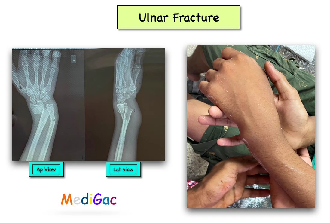
4. Diagnosis :
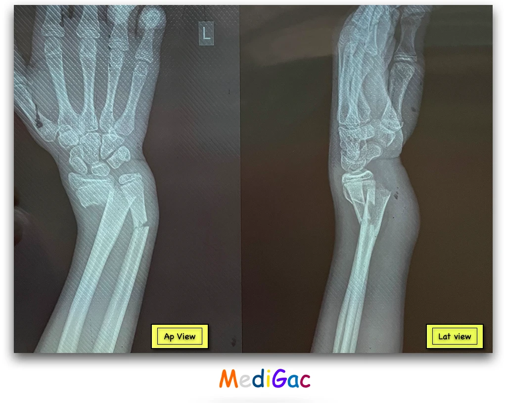
The diagnosis can be based on physical appearance and radiograph.
- Physical appearance : Patient presents with swellings in the forearm region, pain and deformity based on the fracture site.
- Pulse checking – Presence of radial artery pulse means no vascular damage.
- CRT : IF it is <2 seconds that means no vascular injury occurred.
- Finger movements – Suggest no nerve injury.
- Radiograph : We mainly perform Ap and Lateral view xray. Fracture can be seen in the damaged area through X-Ray plate.
5. Treatment :
When patient presents in the emergency ward, we give a pain killer through IM injection, and then send them for X-Ray.
- Conservative treatment – In young/children we can apply plaster cast for six weeks.
- Surgical treatment – In case of adults and older patient we can do various surgeries like locking plate, applying intramedullary nail. If the fracture is displaced we also perform surgery.
5. Complications :
- Malunion in case of conservative treatment.
- Infection.
- Non union.
- Rarely compartment syndrome.