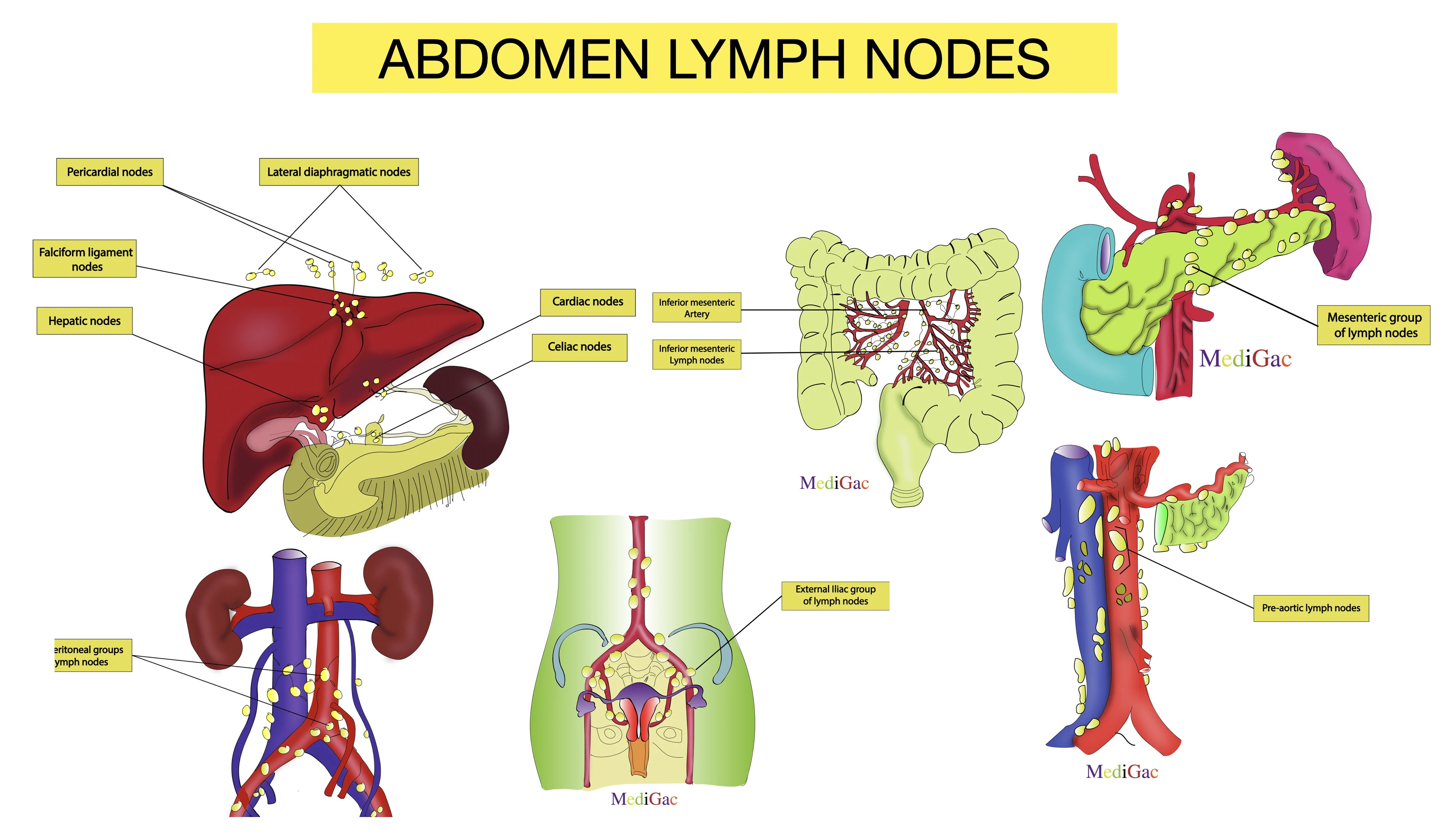
Aortic lymph nodes, Lumbar, Hepatic, Gastric, Splenic and pancreatic, common-internal-external iliac lymph nodes, mesenteric and sacral groups of lymph nodes.
A. Aortic groups of lymph nodes :
1. Lateral aortic groups of lymph nodes :
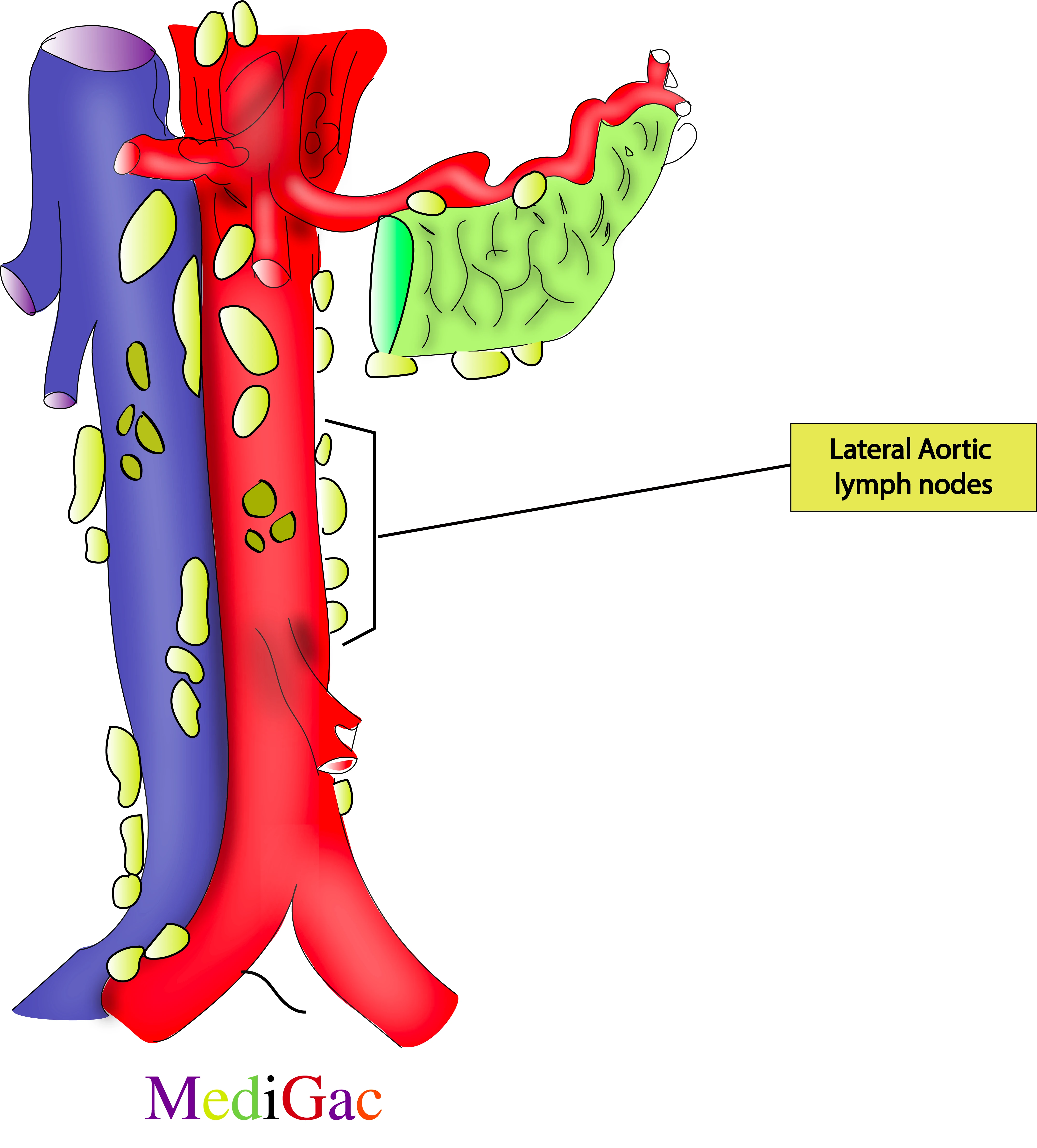
This groups of lymph nodes are present at the lateral side of the abdominal aorta, in between T12 and L4 vertebrae.
I. Location/Position/Relations of lateral aortic lymph nodes :
- Bony relations – Present between L3-L4 vertebra
- Muscle relations – No appropriate muscles around
- Vascular relations – Around the aorta, from where the inferior mesenteric artery originates
- Nervous relations – Sympathetic ganglion lumbar region and Inferior mesenteric ganglion
II. Lymphatic drainage :
- Abdominal lymph empties into the intestinal and lumbar lymphatic trunks, which unite at the level of the second lumbar vertebra to form the cisterna chyli at the base of the thoracic duct, also known as the left lymphatic duct.
- The iliac vessels, the aorta, and the inferior vena cava are the arteries and nodes that carry the lymphatic drainage of the posterior abdominal wall.
- The intestinal and lumbar trunks receive the fluid from these structures, which include the celiac, mesenteric, and iliac nodes.
III. Pathologies of lateral aortic lymph nodes :
- Infection
- GI carcinoma
2. Pre-aortic groups of lymph nodes :
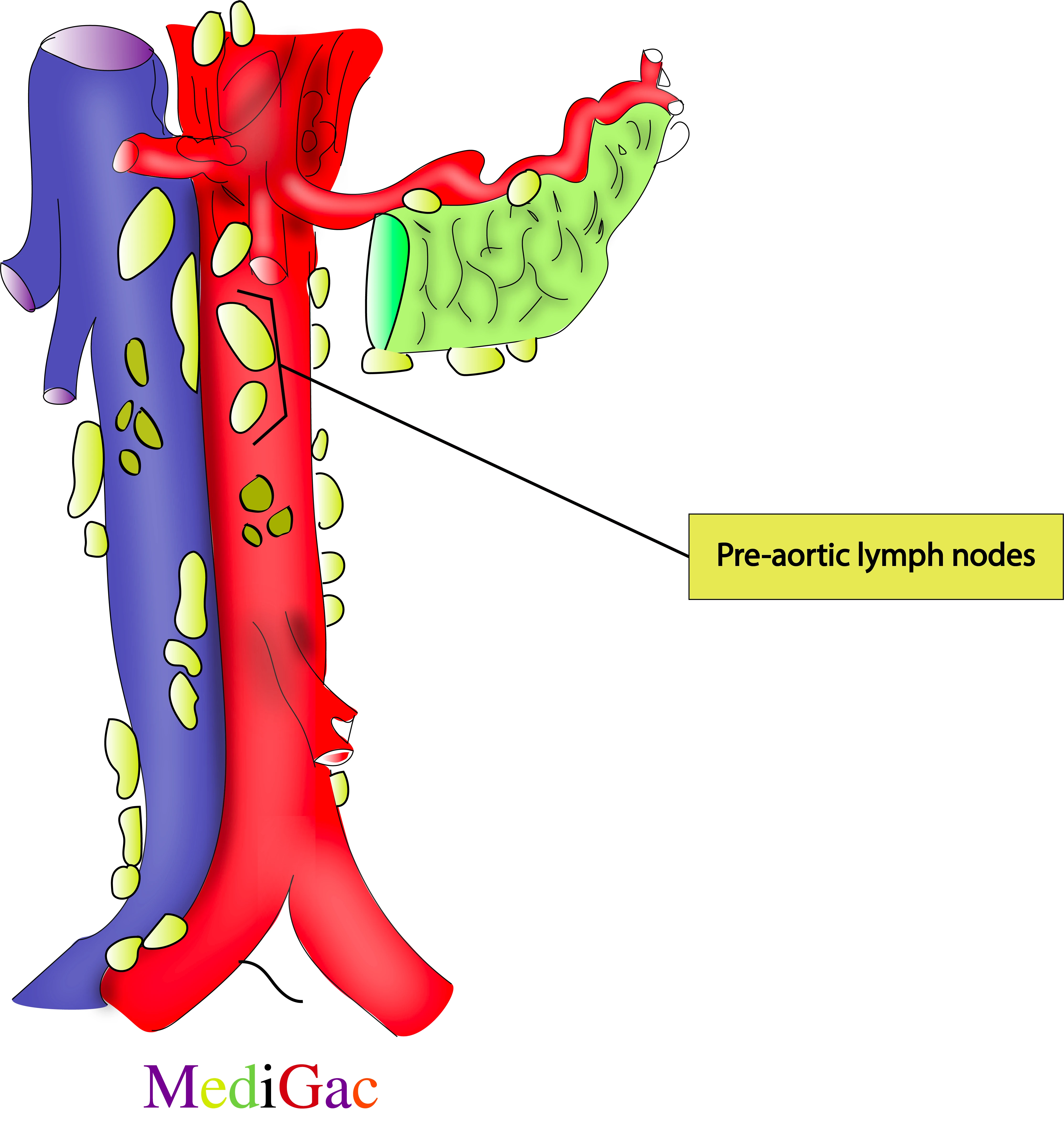
This group of lymph nodes are present in front of the abdominal aorta at the level of L1 vertebrae.
I. Location/Position/Relations of pre-aortic lymph nodes :
- Bony relations – Present between L4-L5 vertebra
- Muscle relations – No appropriate muscle
- Vascular relations – In front of the descending Aorta
- Nervous relations – External Iliac plexus, Aortic plexus and Superior hypogastric plexus
II. Lymphatic drainage :
:
- Abdominal lymph empties into the intestinal and lumbar lymphatic trunks, which unite at the level of the second lumbar vertebra to form the cisterna chyli at the base of the thoracic duct, also known as the left lymphatic duct.
- The iliac vessels, the aorta, and the inferior vena cava are the arteries and nodes that carry the lymphatic drainage of the posterior abdominal wall.
- The intestinal and lumbar trunks receive the fluid from these structures, which include the celiac, mesenteric, and iliac nodes.
III. Pathologies of Pre-aortic lymph nodes :
- Infections
- Acute phase of Kawasaki disease
- Cancer
3. Retro aortic or post aortic groups of lymph nodes :
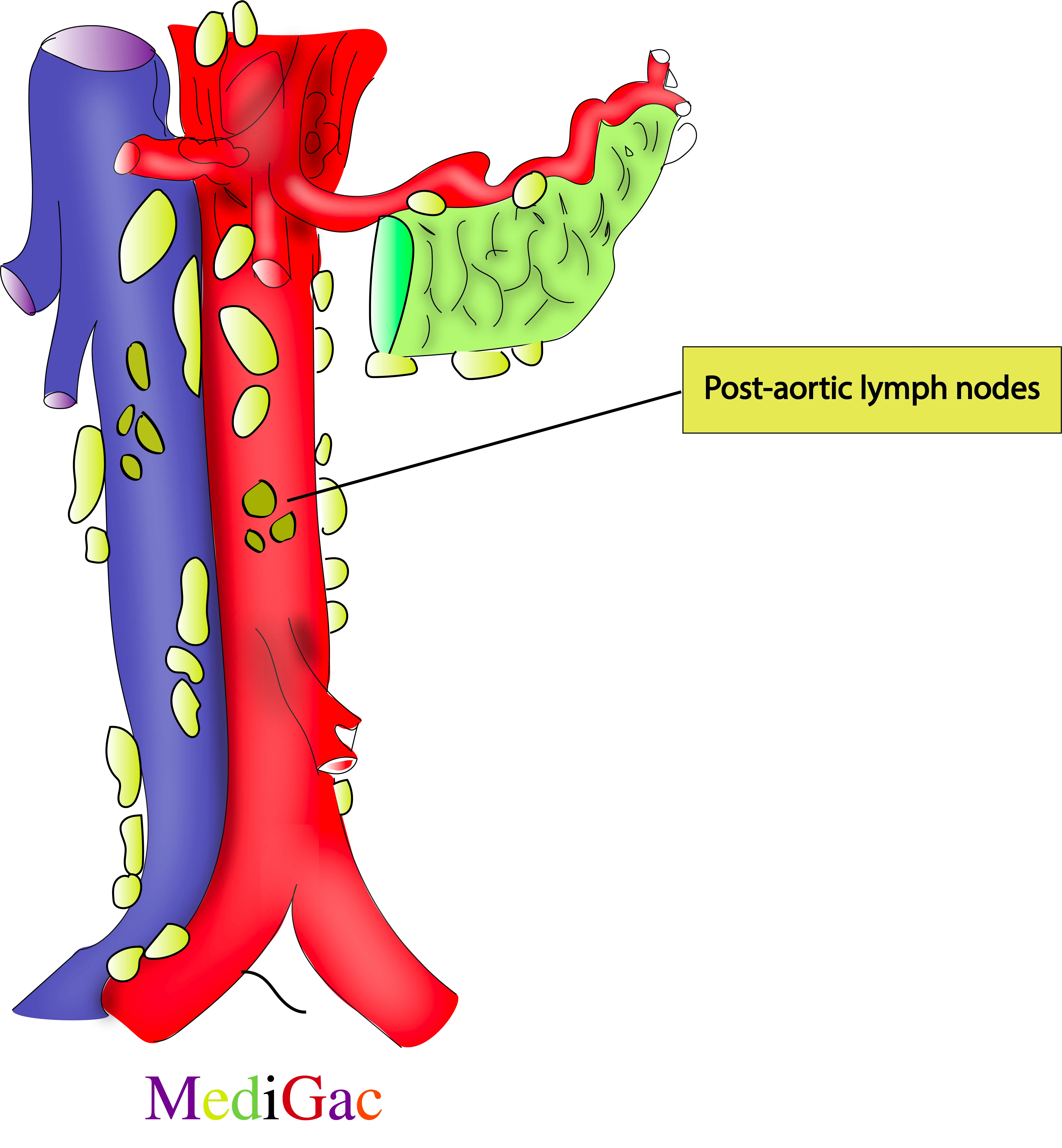
This group of lymph nodes are present behind the abdominal aorta at the level of L3-L4 vertebrae.
I. Location/Position/Relations of post-aortic lymph nodes :
- Bony relations – Present in front of L3 vertebra
- Muscle relations – No appropriate muscle
- Vascular relations – Back of descending Aorta
- Nervous relations – Inferior mesenteric ganglion
II. Lymphatic drainage :
- Abdominal lymph empties into the intestinal and lumbar lymphatic trunks, which unite at the level of the second lumbar vertebra to form the cisterna chyli at the base of the thoracic duct, also known as the left lymphatic duct.
- The iliac vessels, the aorta, and the inferior vena cava are the arteries and nodes that carry the lymphatic drainage of the posterior abdominal wall.
- The intestinal and lumbar trunks receive the fluid from these structures, which include the celiac, mesenteric, and iliac nodes.
III. Pathologies of post-aortic lymph nodes :
- Infections such as tuberculosis
- Cancer
- Inflammatory conditions such as sarcoidosis
- Blood cancer
B. Lumbar groups of lymph nodes :
1.Intermediate groups of lymph nodes :
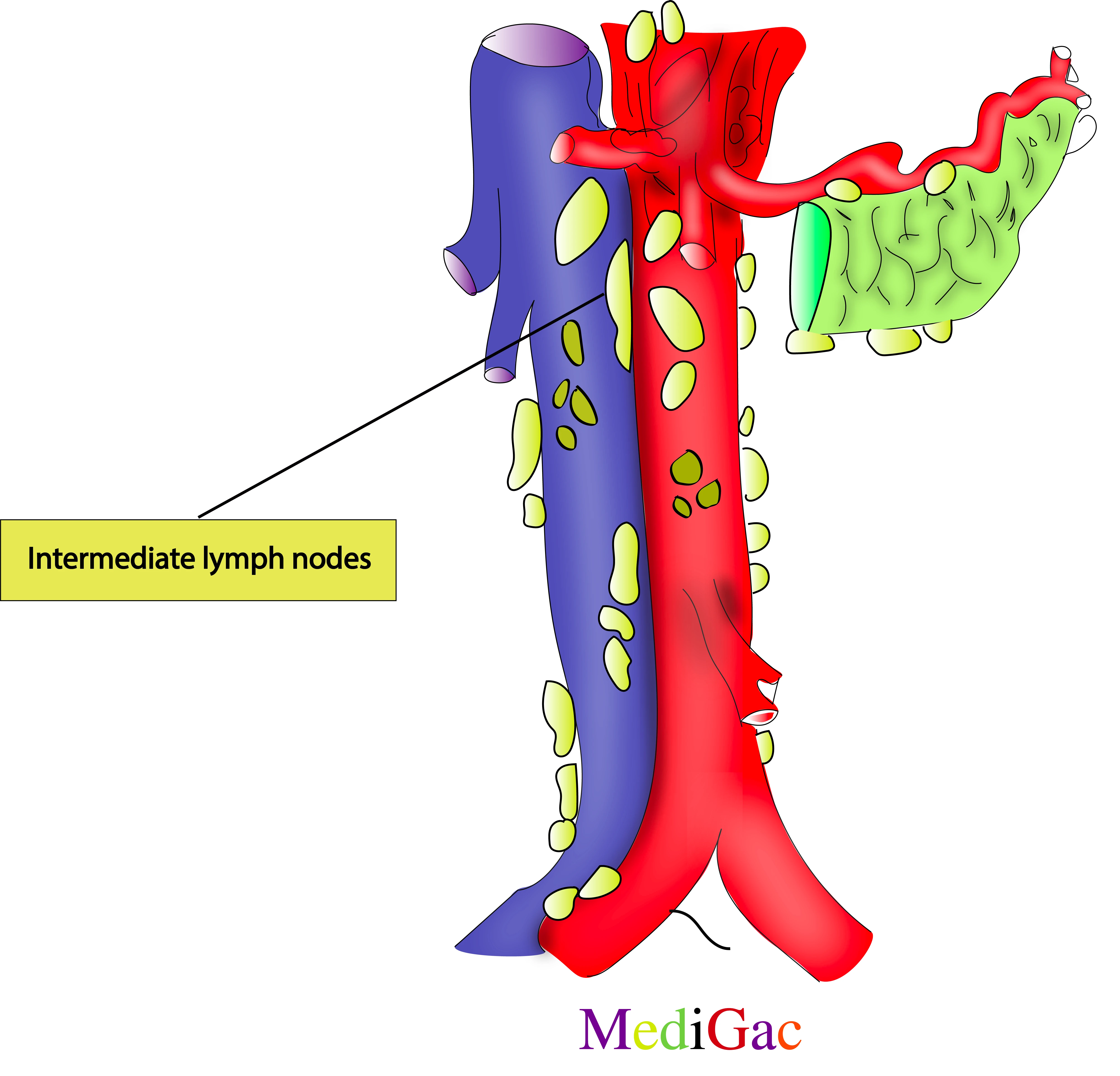
These are present at the right side and in front of the abdominal aorta through out the lumbar vertebrae that is from L1 to L5.
I. Location/Position/Relations of Intermediate lymph nodes :
- Bony relations – Presents along the L3-L5 vertebra
- Muscle relations – No appropriate muscles
- Vascular relations – Along the right side of the descending aorta
- Nervous relations – Intermesenteric plexus
II. Lymphatic drainage :
- Abdominal lymph empties into the intestinal and lumbar lymphatic trunks, which unite at the level of the second lumbar vertebra to form the cisterna chyli at the base of the thoracic duct, also known as the left lymphatic duct.
- The iliac vessels, the aorta, and the inferior vena cava are the arteries and nodes that carry the lymphatic drainage of the posterior abdominal wall.
- The intestinal and lumbar trunks receive the fluid from these structures, which include the celiac, mesenteric, and iliac nodes.
III. Pathologies of Intermediate lymph nodes :
- Ovarian cancer
- Acute phase of Kawasaki disease
2. Lateral caval groups of lymph nodes :
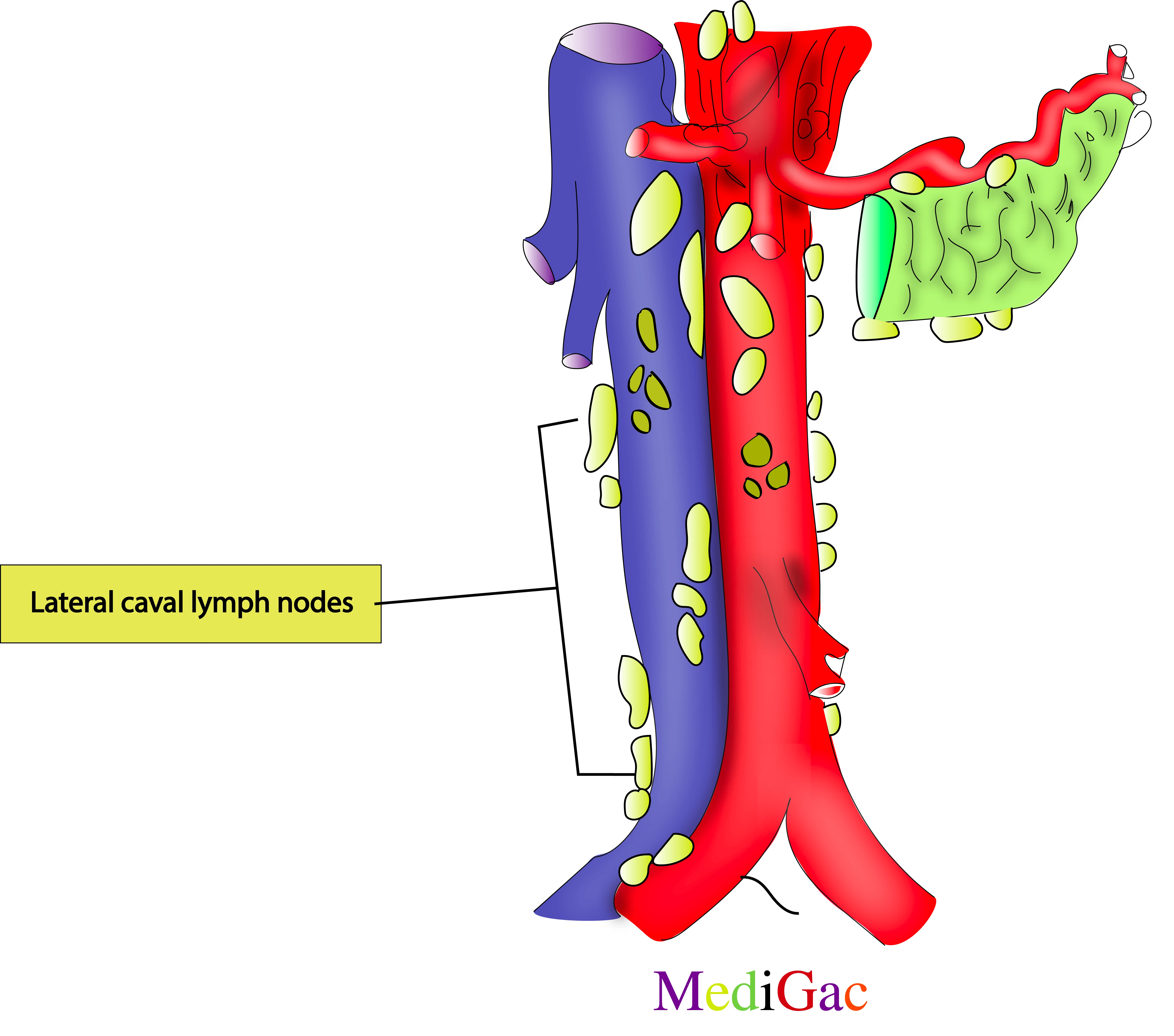
Normally canal means veins, lateral canal signifies lateral side of inferior vena cava. So this groups of lymph nodes are present lateral side if the IVC at the level of
I. Location/Position/Relations of Lateral caval lymph nodes :
- Bony relations – Along the L1-L5 vertebra
- Muscle relations – No appropriate muscle
- Vascular relations – Along the right side of the inferior vena cava
- Nervous relations – Right sided sympathetic trunk
II. Lymphatic drainage :
- Abdominal lymph empties into the intestinal and lumbar lymphatic trunks, which unite at the level of the second lumbar vertebra to form the cisterna chyli at the base of the thoracic duct, also known as the left lymphatic duct.
- The iliac vessels, the aorta, and the inferior vena cava are the arteries and nodes that carry the lymphatic drainage of the posterior abdominal wall.
- The intestinal and lumbar trunks receive the fluid from these structures, which include the celiac, mesenteric, and iliac nodes.
III. Pathologies of Lateral caval lymph nodes :
- GI cancer
- Peritoneal abscess
- Infection
L1-L5 vertebrae that is through out the lumbar vertebrae.
3. Precaval groups of lymph nodes :
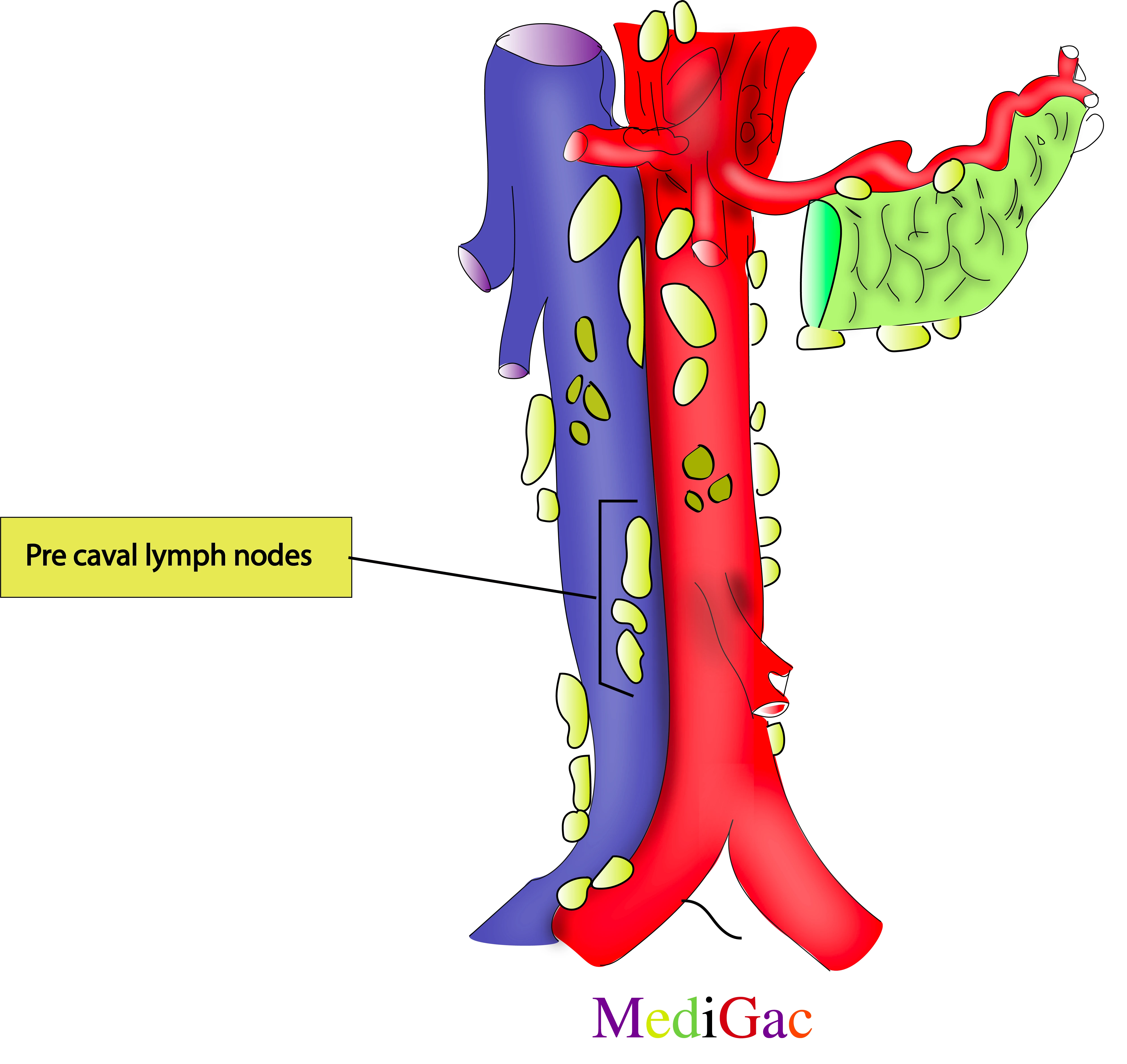
These are situated just in front of the IVC at the level of L3 to L4 vertebrae.
I. Location/Position/Relations of Pre-caval lymph nodes :
- Bony relations – Along the L3-L4 vertebra
- Muscle relations – No such
- Vascular relations – Back of the Inferior vena cava
- Nervous relations – Sympathetic trunk
II. Lymphatic drainage :
- Abdominal lymph empties into the intestinal and lumbar lymphatic trunks, which unite at the level of the second lumbar vertebra to form the cisterna chyli at the base of the thoracic duct, also known as the left lymphatic duct.
- The iliac vessels, the aorta, and the inferior vena cava are the arteries and nodes that carry the lymphatic drainage of the posterior abdominal wall.
- The intestinal and lumbar trunks receive the fluid from these structures, which include the celiac, mesenteric, and iliac nodes.
III. Pathologies of Pre-caval lymph nodes :
- Colon Carcinoma
- Infection from bacteria or viruses
4. Postcaval/Retrocaval groups of lymph nodes :
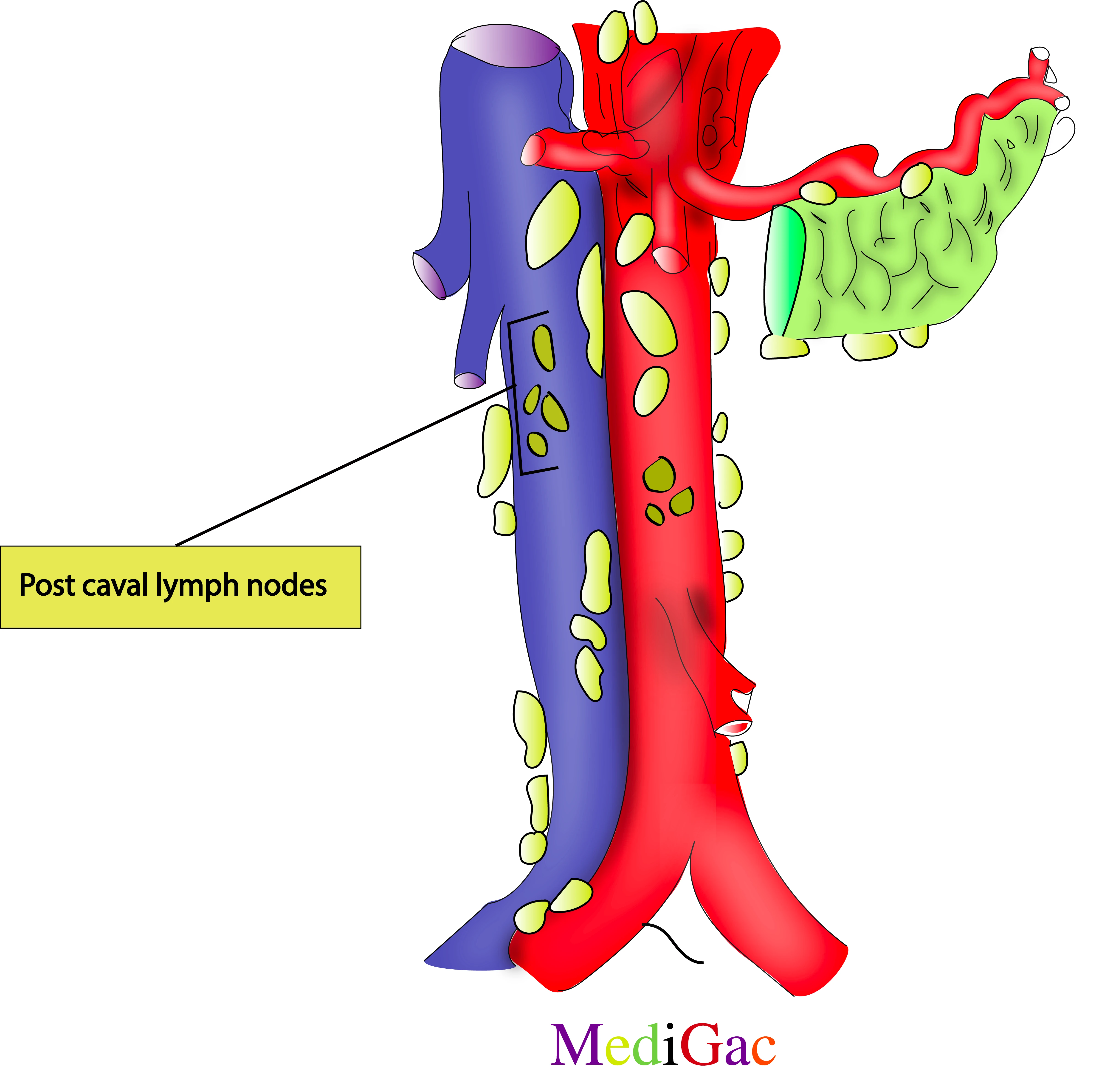
These lymph nodes are present behind the IVC at the level of L1-L5 vertebrae.
I. Location/Position/Relations of Retro-caval lymph nodes :
- Bony relations – L1-L5 vertebra
- Muscle relations – No such
- Vascular relations – Inferior vena cava
- Nervous relations – Sympathetic trunk
II. Lymphatic drainage :
- Abdominal lymph empties into the intestinal and lumbar lymphatic trunks, which unite at the level of the second lumbar vertebra to form the cisterna chyli at the base of the thoracic duct, also known as the left lymphatic duct.
- The iliac vessels, the aorta, and the inferior vena cava are the arteries and nodes that carry the lymphatic drainage of the posterior abdominal wall.
- The intestinal and lumbar trunks receive the fluid from these structures, which include the celiac, mesenteric, and iliac nodes.
III. Pathologies of Retro-caval lymph nodes :
- Infection from bacteria and viruses
- Cancer
C. Visceral groups of lymph nodes :
1.Hepatic groups of lymph nodes :
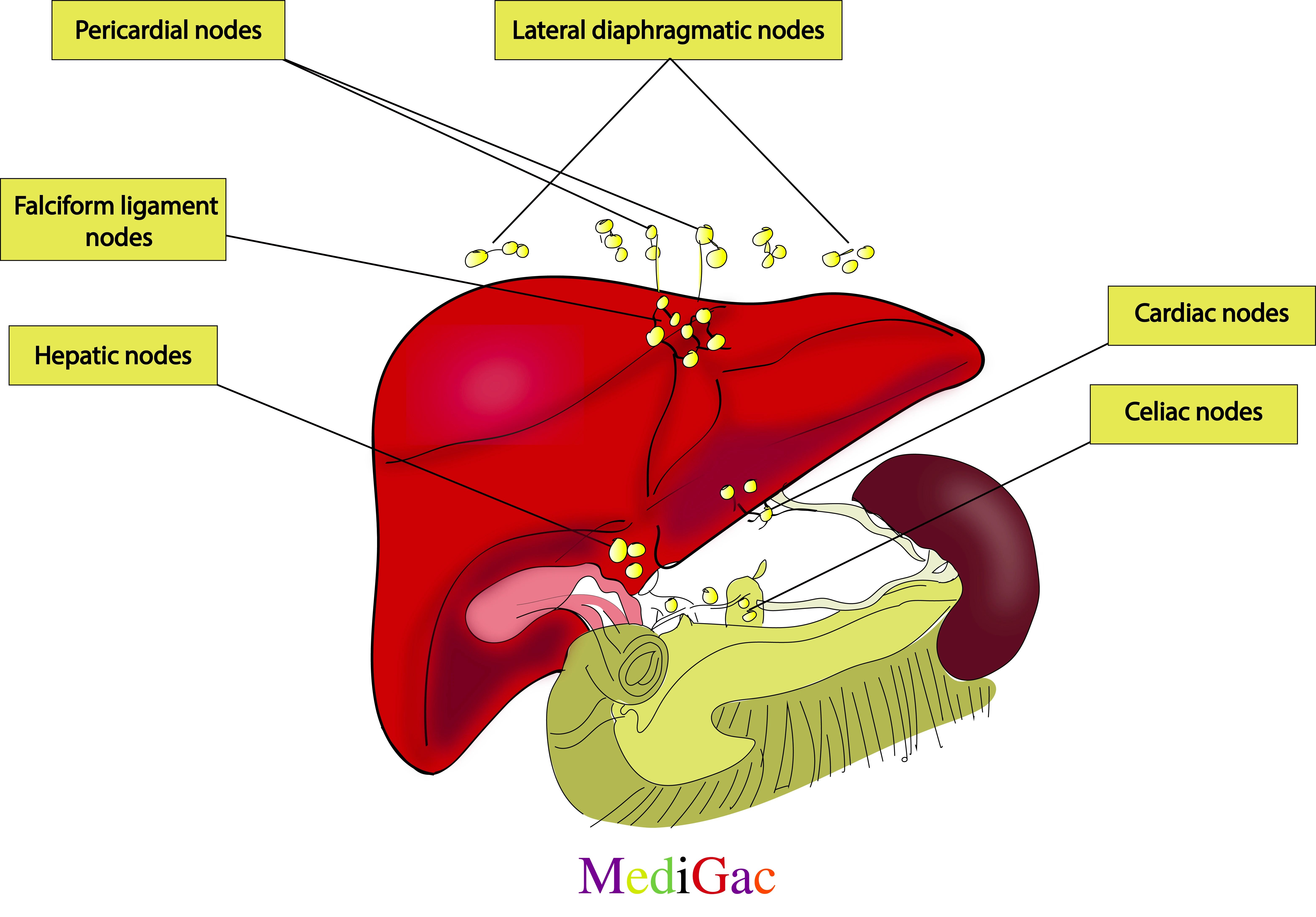
- Hepatic nodes – These nodes are present along the common hepatic duct.
- Celiac nodes – These are also pre aortic lymph nodes, because the presence of this nodes are infant of the abdominal aorta. Present along the celiac trunk between T12 and L1 vertebrae.
- Pericardial nodes – These groups of lymph nodes are present at the level of T7-T9 vertebrae.
- Falciform nodes – These are present along the falciform ligament.
- Cardiac/Juxtaesophageal(right and left) : Right – Present at the right side of the gastroesophageal junction and Left : Present at the left side of the lower part of the esophagus.
II. Lymphatic drainage :
- Many clusters of lymph nodes surround the liver, which generates up to half of the body’s lymph.
- The hepatic, left gastric, mediastinal, cystic, phrenic, and celiac nodes are among these groupings.
- The cisternachyli receives lymph that drains from the hepatic nodes into the celiac nodes. The right lymphatic duct is joined by additional hepatic veins.
III. Pathologies of Hepatic lymph nodes :
- Chronic hepatitis B
- Cholangiocarcinoma
- Galbladder carcinoma
- Obstruction of the bile duct at the level of the hilum of the liver
2. Gastric groups of lymph nodes :
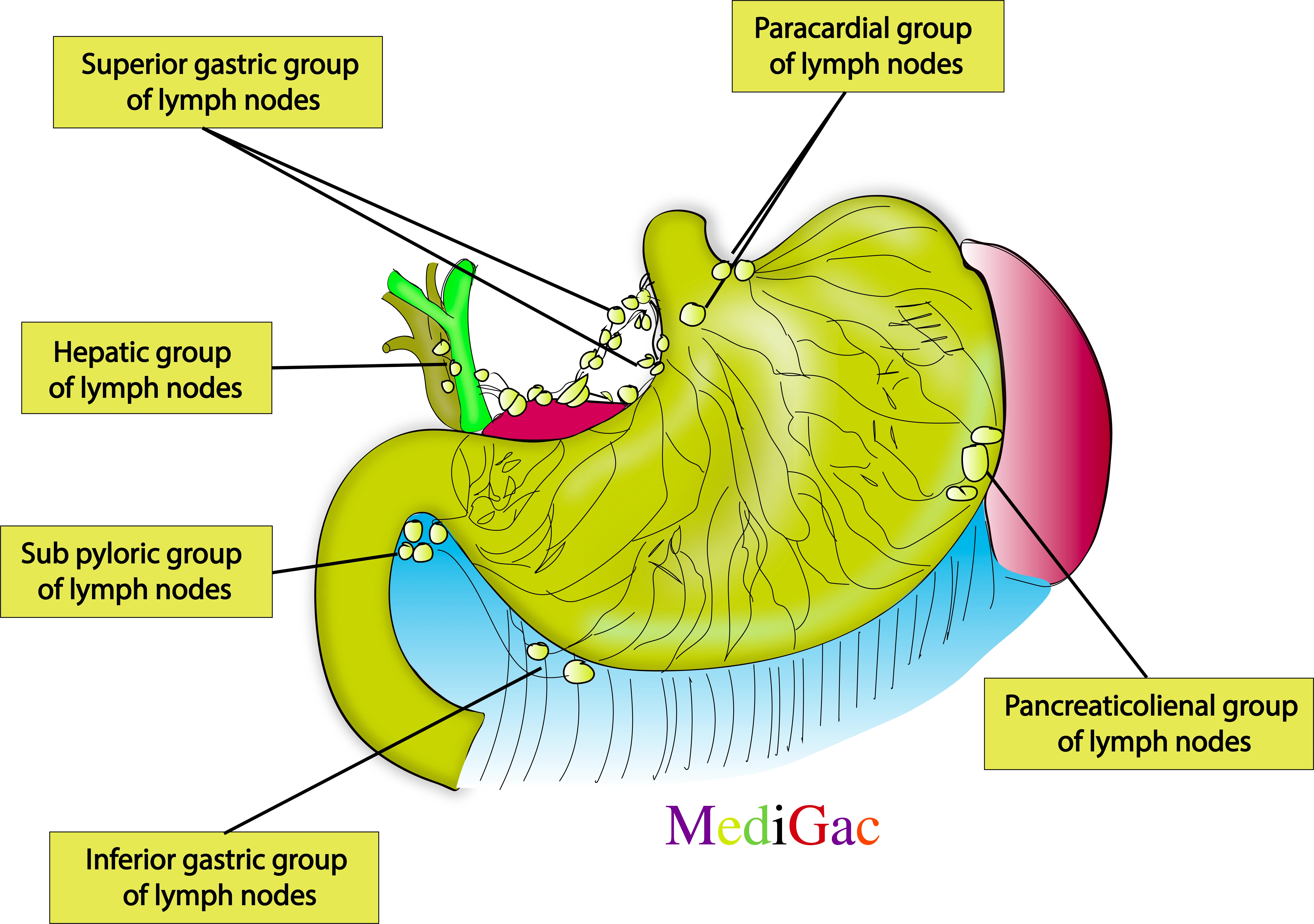
- Superior gastric lymph nodes – These are present around the lesser curvature of the stomach.
- Pericardial – Present along the cardiac part of the stomach.
- Sub-pyloric – These are present below the pyloric end of the stomach.
- Inferior gastric – These nodes are present at the inferior part of the greater curvature of stomach.
- Pancreaticolienal – These includes pancreaticosplenic and superior pancreatic lymph nodes.
II. Lymphatic drainage :
The majority of the stomach’s lymphatic outflow passes through intermediary nodes before arriving at the celiac nodes. The lymph flowing from the stomach wall empties into lymphatic vessels that start in the mucosa, develop into a dense network in the submucosa, and finally converge at the sub-peritoneal plexus.
III. Pathologies of Gastric lymph nodes :
- Infection from bacteria and viruses
- Gastroenteritis
3. Splenic groups of lymph nodes :
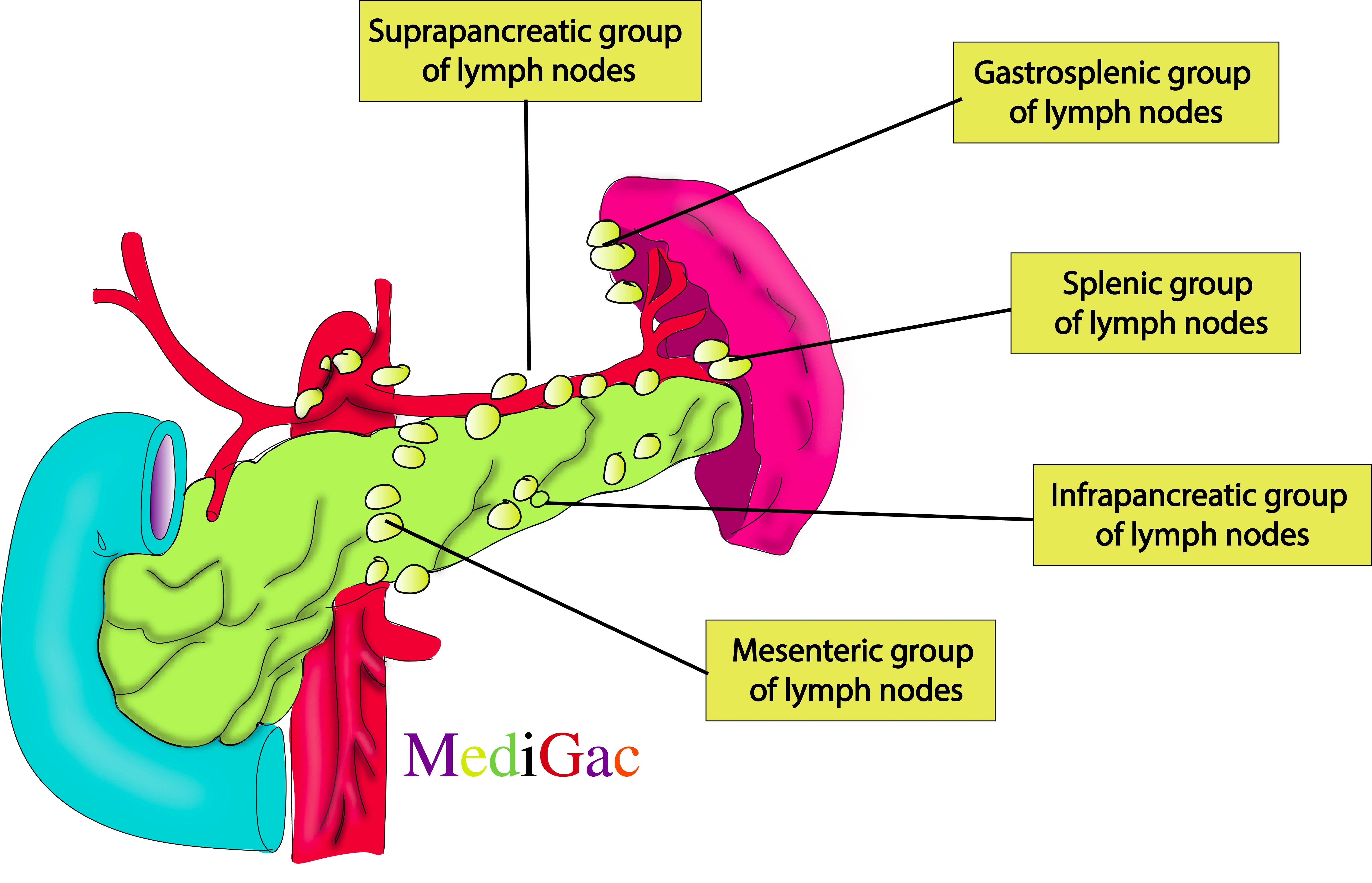
- Superior pancreatic – These are present at the superior border of pancreas.
- Gastrosplenic – Present along the gastrosplenic ligament.
- Splenic – Present along the insertion of splenic vessels into the spleen.
- Infra pancreatic – These are present along the inferior border of the pancreas.
- Mesenteric – Present along the mesentery.
II. Lymphatic drainage :
- Abdominal lymph empties into the intestinal and lumbar lymphatic trunks, which unite at the level of the second lumbar vertebra to form the cisterna chyli at the base of the thoracic duct, also known as the left lymphatic duct.
- The iliac vessels, the aorta, and the inferior vena cava are the arteries and nodes that carry the lymphatic drainage of the posterior abdominal wall.
- The intestinal and lumbar trunks receive the fluid from these structures, which include the celiac, mesenteric, and iliac nodes.
III. Pathologies of Splenic lymph nodes :
- Infection
- Cancer
D. Groups of lymph nodes around Iliac vessels :
1. Common iliac groups of lymph nodes :
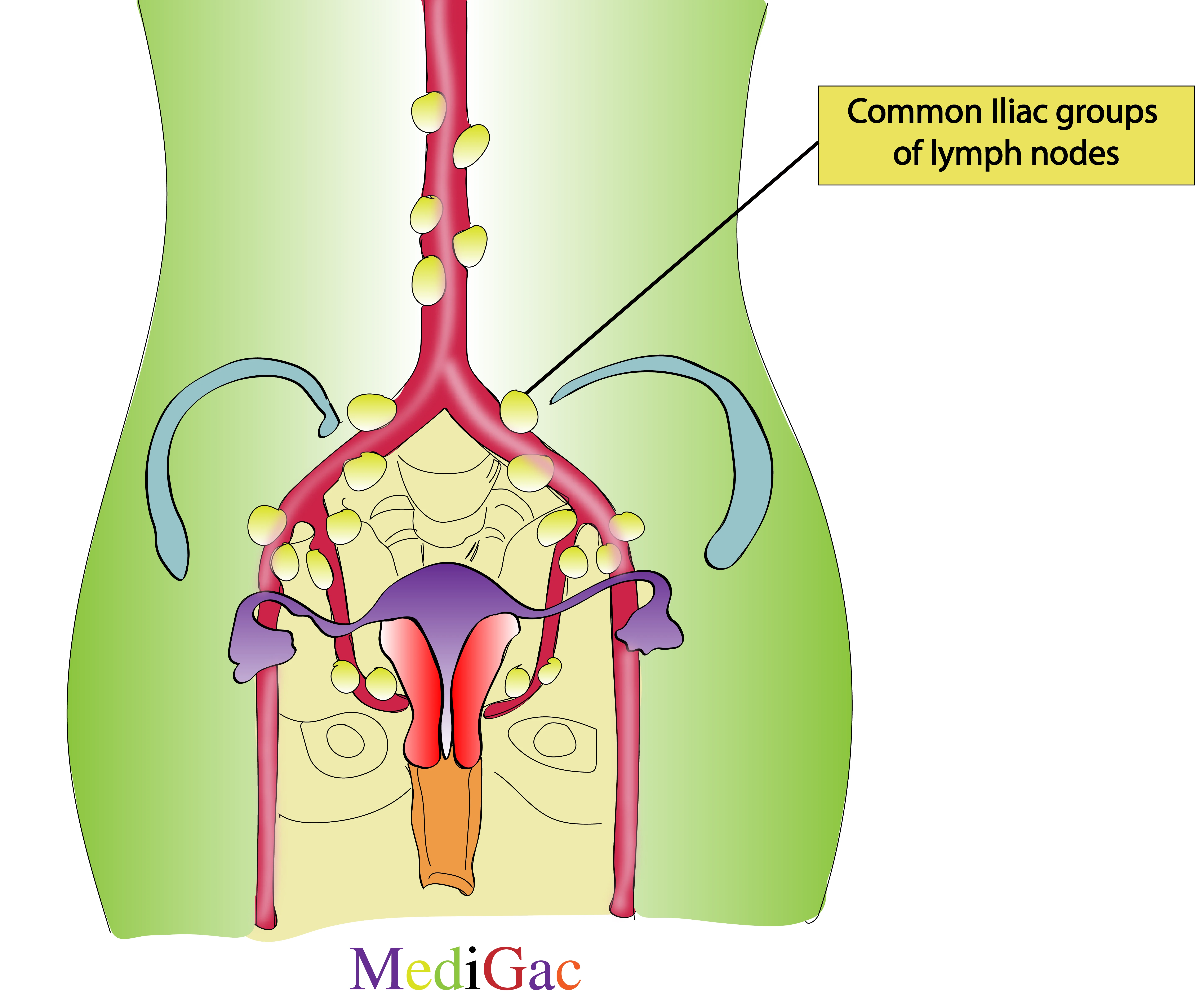
These lymph nodes are presents around the common iliac artery.
I. Location/Position/Relations of Common Iliac lymph nodes :
- Bony relations – L5 vertebra
- Muscle relations – No such
- Vascular relations – Common iliac artery
- Nervous relations – External iliac plexus
II. Lymphatic drainage :
The common iliac lymph nodes, which are located beneath the abdominal lumbar nodes, receive lymph from the external and internal iliac lymph nodes. Lymph from the pelvic region is transported by the lumbar and common iliac nodes into the lumbar trunks and then into the cisterna chyli at the base of the thoracic duct, also known as the left lymphatic duct.
III. Pathologies of Common Iliac lymph nodes :
- Cancer of cervix
- Skin infection like Cellulitis
- Infection from bacteria and viruses
2. Internal iliac groups of lymph nodes :
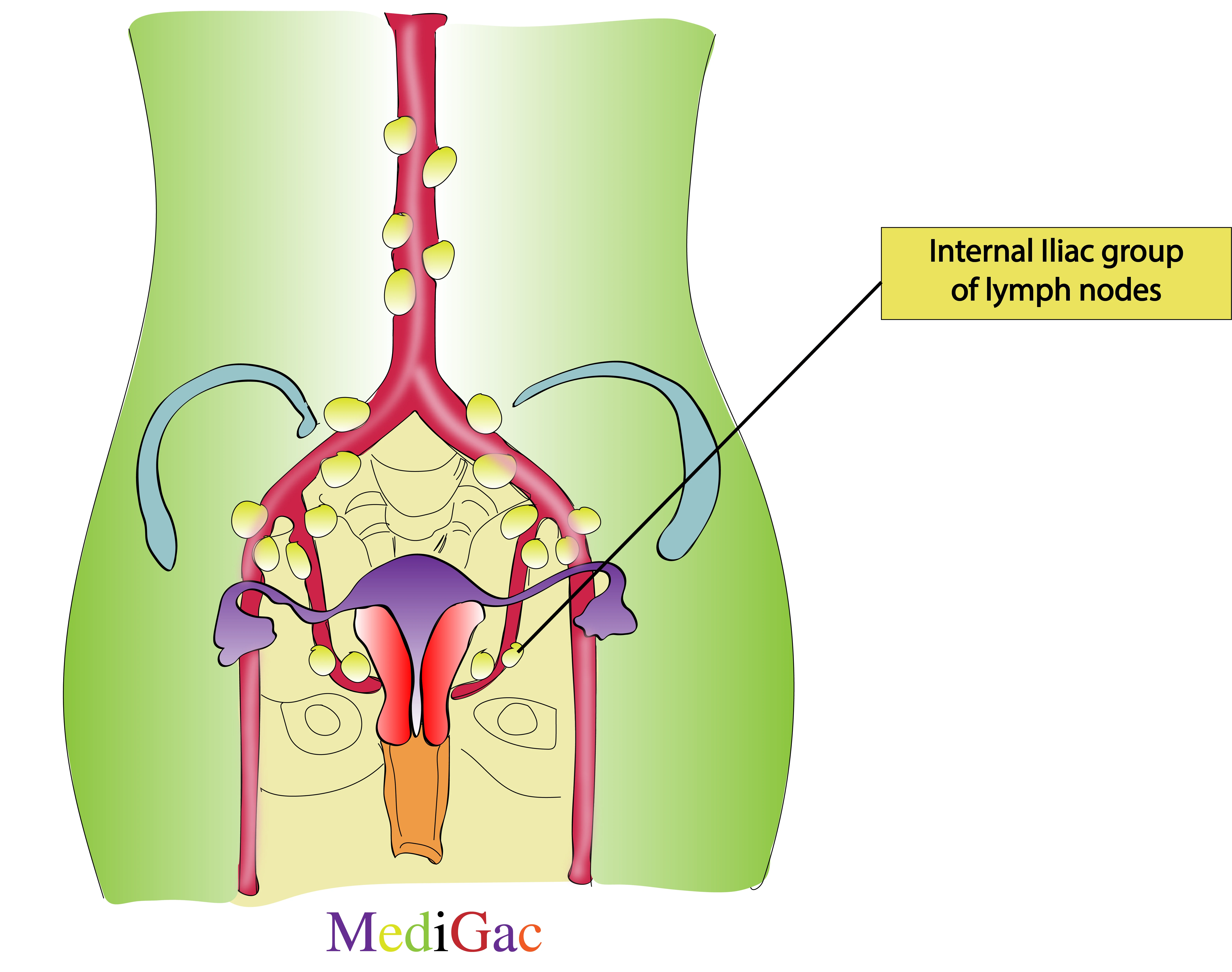
Present along the internal iliac artery.
I. Location/Position/Relations of Internal Iliac lymph nodes :
- Bony relations – Sacrum S1
- Muscle relations – No such
- Vascular relations – Internal iliac artery
- Nervous relations – Sacral ganglion
II. Lymphatic drainage :
The common iliac lymph nodes, which are located beneath the abdominal lumbar nodes, receive lymph from the external and internal iliac lymph nodes. Lymph from the pelvic region is transported by the lumbar and common iliac nodes into the lumbar trunks and then into the cisterna chyli at the base of the thoracic duct, also known as the left lymphatic duct.
III. Pathologies of Internal Iliac lymph nodes :
- Infection from Bactria and viruses
- Malignancy
3. External iliac groups of lymph nodes :
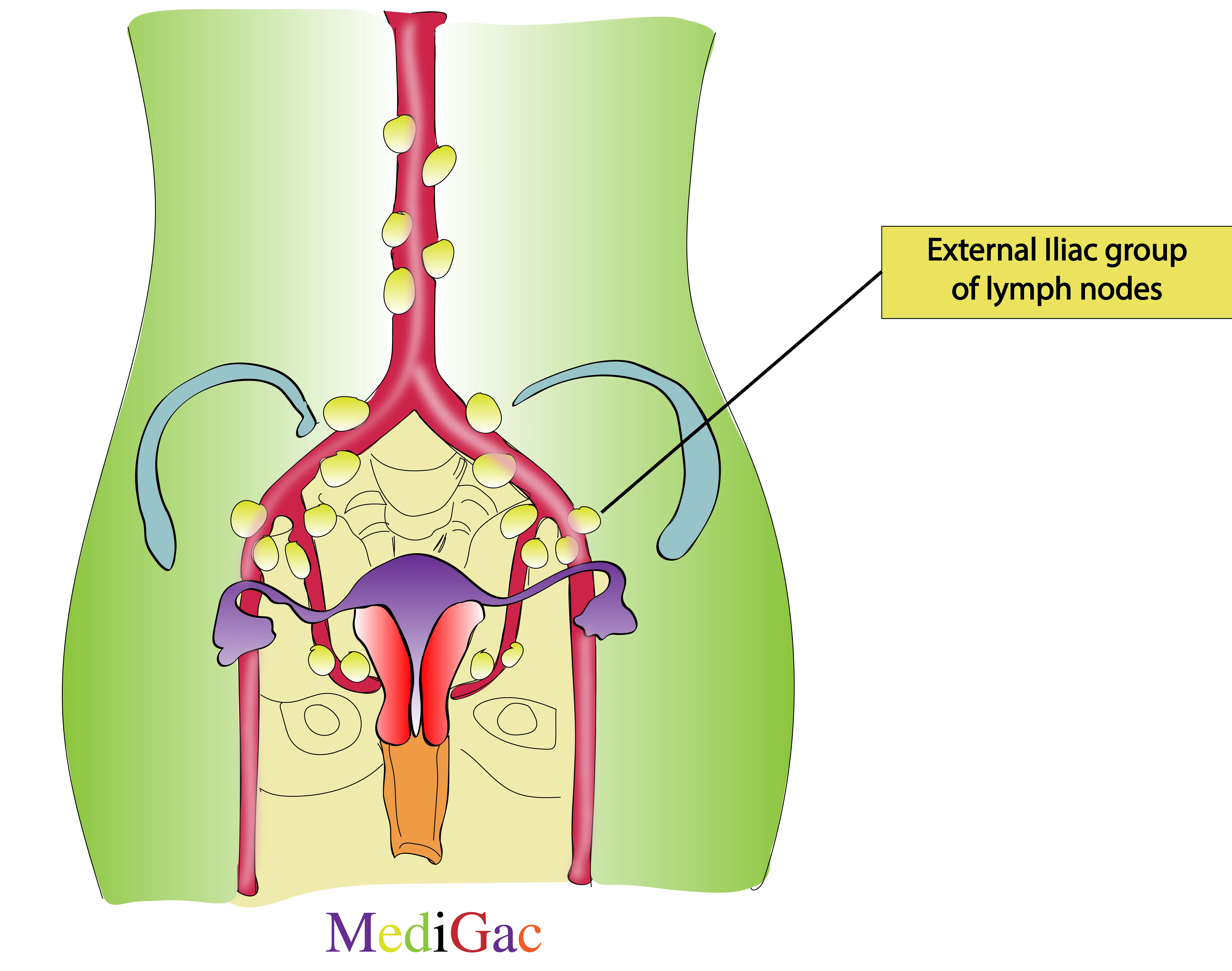
Present along the external iliac artery.
I. Location/Position/Relations of External Iliac lymph nodes :
- Bony relations – Sacrum S1-S5
- Muscle relations – Psoas major middle portion
- Vascular relations – External Iliac artery
- Nervous relations – Genitofemoral nerve
II. Lymphatic drainage :
The common iliac lymph nodes, which are located beneath the abdominal lumbar nodes, receive lymph from the external and internal iliac lymph nodes. Lymph from the pelvic region is transported by the lumbar and common iliac nodes into the lumbar trunks and then into the cisterna chyli at the base of the thoracic duct, also known as the left lymphatic duct.
III. Pathologies of External Iliac lymph nodes :
- Infection from Bactria and viruses
- Malignancy
E. Mesenteric groups of lymph nodes :
1. Superior mesenteric groups of lymph nodes :
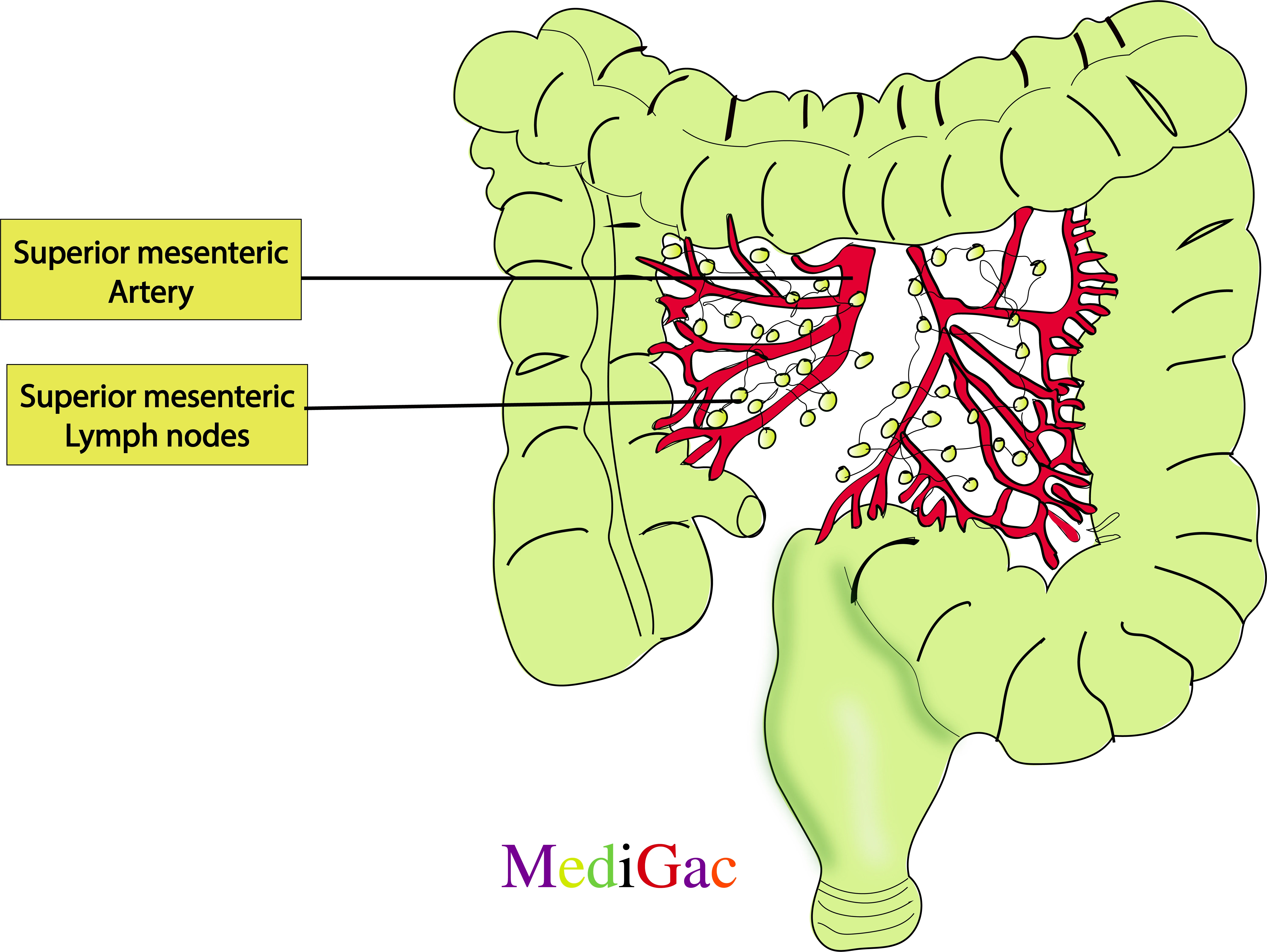
These lymph nodes are present surrounding the superior mesenteric artery.
I. Location/Position/Relations of Superior mesenteric lymph nodes :
- Bony relations – T12-L2 Vertebra
- Muscle relations – No such
- Vascular relations – Along the superior mesenteric artery
- Nervous relations – Superior mesenteric plexus
II. Lymphatic drainage :
- Abdominal lymph empties into the intestinal and lumbar lymphatic trunks, which unite at the level of the second lumbar vertebra to form the cisterna chyli at the base of the thoracic duct, also known as the left lymphatic duct.
- The iliac vessels, the aorta, and the inferior vena cava are the arteries and nodes that carry the lymphatic drainage of the posterior abdominal wall.
- The intestinal and lumbar trunks receive the fluid from these structures, which include the celiac, mesenteric, and iliac nodes.
III. Pathologies of Superior mesenteric lymph nodes :
- Non Neoplastic causes : Appendicitis, Chron’s disease, Procto colitis
- Neoplastic causes : Lymphoma, Colonic cancer, Breast carcinoma, Gastrointestinal carcinoid tumour, Adenocarcinoma of the pancreas
- Infection : Tuberculosis
2. Inferior mesenteric groups of lymph nodes :
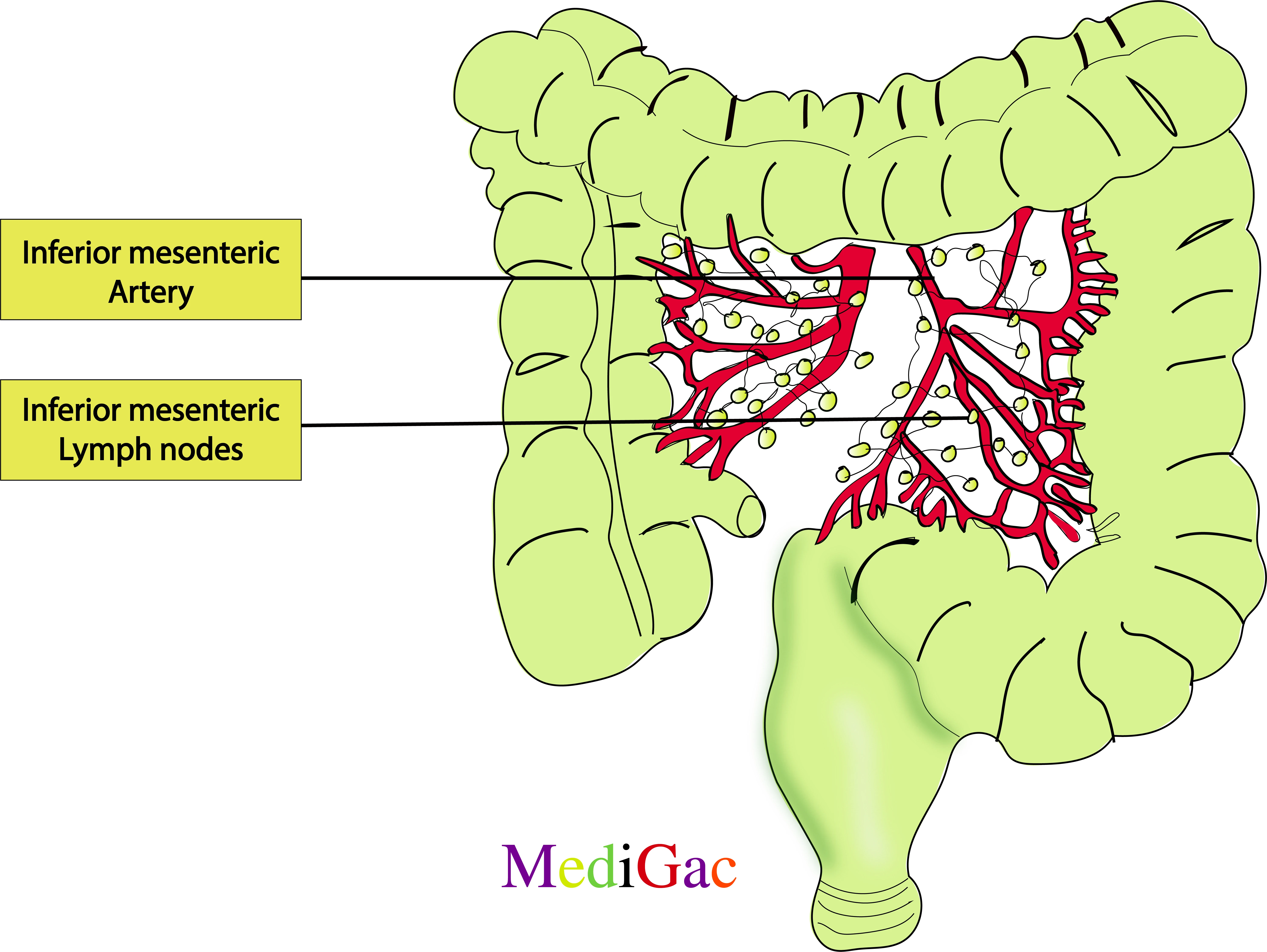
These are present surrounding the inferior mesenteric artery.
I. Location/Position/Relations of Inferior mesenteric lymph nodes :
- Bony relations – L3 vertebra
- Muscle relations – No such
- Vascular relations – Inferior mesenteric artery
- Nervous relations – Inferior mesenteric ganglion
II. Lymphatic drainage :
- Abdominal lymph empties into the intestinal and lumbar lymphatic trunks, which unite at the level of the second lumbar vertebra to form the cisterna chyli at the base of the thoracic duct, also known as the left lymphatic duct.
- The iliac vessels, the aorta, and the inferior vena cava are the arteries and nodes that carry the lymphatic drainage of the posterior abdominal wall.
- The intestinal and lumbar trunks receive the fluid from these structures, which include the celiac, mesenteric, and iliac nodes.
III. Pathologies of Inferior mesenteric lymph nodes :
- Non Neoplastic causes : Appendicitis, Chron’s disease, Procto colitis
- Neoplastic causes : Lymphoma, Colonic cancer, Breast carcinoma, Gastrointestinal carcinoid tumour, Adenocarcinoma of the pancreas
- Infection : Tuberculosis
F. Others groups of lymph nodes :
1. Sacral groups of lymph nodes :
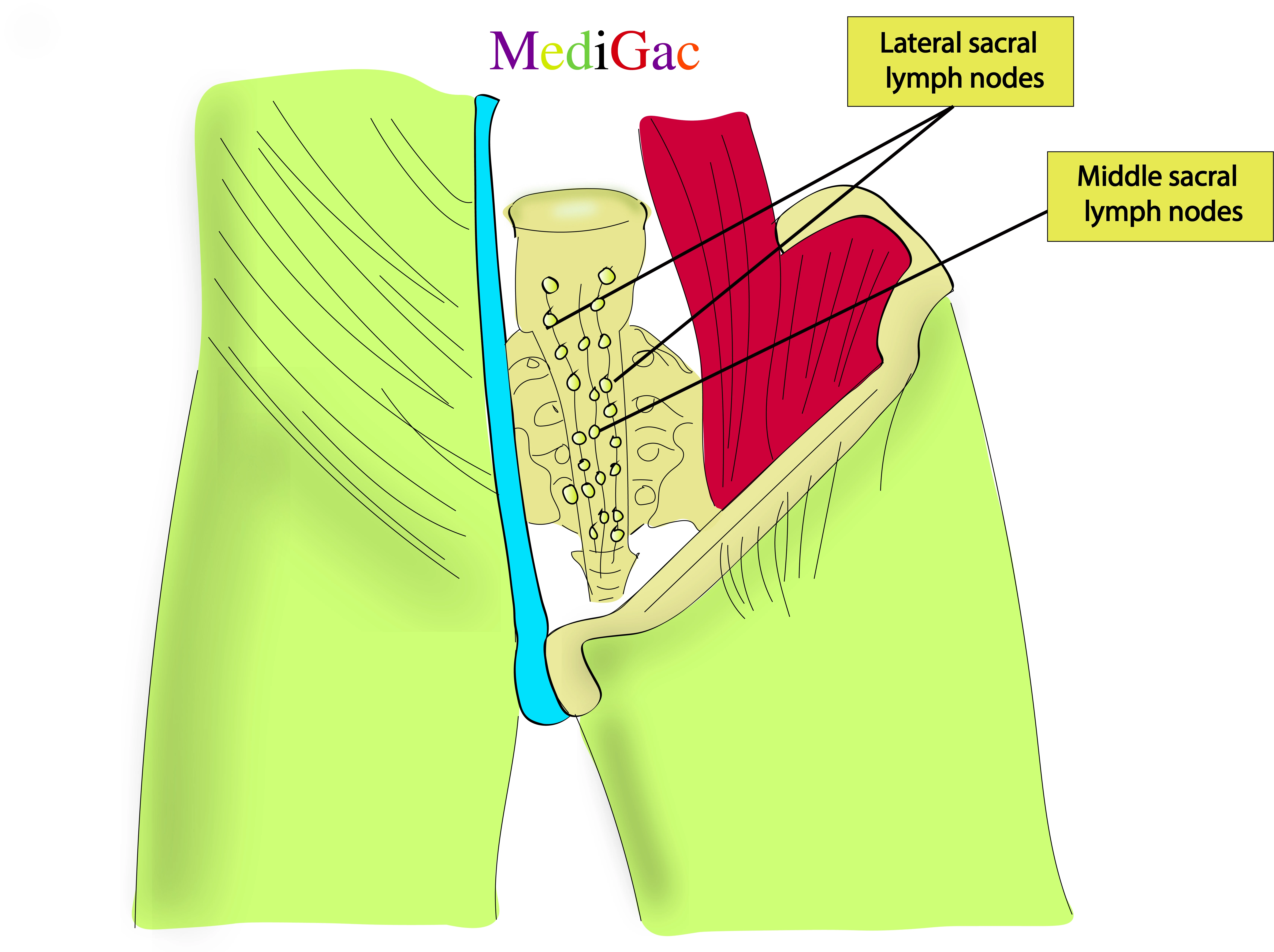
- Lateral sacral – Along the lateral border of the sacrum.
- Middle sacral – Along the middle border of the sacrum.
Pathology :
- Infection from bacteria or viruses
- Hodgkin lymphoma
2. Retro-peritoneal groups of lymph nodes :

This group of lymph nodes are present behind the abdominal aorta at the level of L3-L4 vertebrae.
I. Location/Position/Relations of Retroperitoneal lymph nodes :
- Bony relations – L3-L4 Vertebra
- Muscle relations – Behind the Anterior longitudinal ligament
- Vascular relations – Descending aorta
- Nervous relations – Intermesenteric plexus
II. Lymphatic drainage :
- The iliac vessels, the aorta, and the inferior vena cava are the arteries and nodes that carry the lymphatic drainage of the posterior abdominal wall.
- The intestinal and lumbar trunks receive the fluid from these structures, which include the celiac, mesenteric, and iliac nodes.
III. Pathologies of Retroperitoneal lymph nodes :
- Cancer spread to lymph nodes
- Infections like tuberculosis
- Non cancerous growth like Castleman disease
- Testicular cancer
- Hodgkin lymphoma nad Non-hodgkin lymphoma